Tang Qiying, Liu Xinyou, Jiang Qiuli, Zhu Liuhong, Zhang Jinhui, Wu Pu-Yeh, Jiang Ying, Zhou Jianjun
Department of Radiology, Xiamen Branch, Zhongshan Hospital, Fudan University, Xiamen, China.
Department of General Surgery, Xiamen Branch, Zhongshan Hospital, Fudan University, Xiamen, China.
Quant Imaging Med Surg. 2023 Apr 1;13(4):2697-2707. doi: 10.21037/qims-22-172. Epub 2022 Aug 18.
The aim of this study was to investigate the value of unenhanced magnetic resonance imaging (MRI) with diffusion kurtosis imaging (DKI) in diagnosing papillary thyroid carcinoma (PTC).
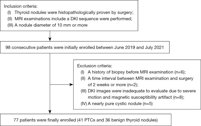
In all, 77 consecutive patients comprising a total of 77 thyroid nodules were enrolled in this study. Of these nodules, 41 were histopathologically confirmed PTCs and 36 were benign nodules. All patients underwent thyroid MRI including T1-weighted imaging (T1WI), T2-weighted imaging (T2WI), diffusion-weighted imaging (DWI), and DKI. All the images were assessed by 2 radiologists. The signal intensity ratio (SIR) of these nodules on T1WI and T2WI, the apparent diffusion coefficient (ADC) from DWI, and mean diffusivity (MD) and mean kurtosis (MK) from DKI were measured. Morphological features on these images were also evaluated. Univariate and multivariate logistic regression analyses were used to evaluate the value of these parameters as potential predictors of PTC.
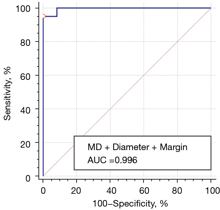
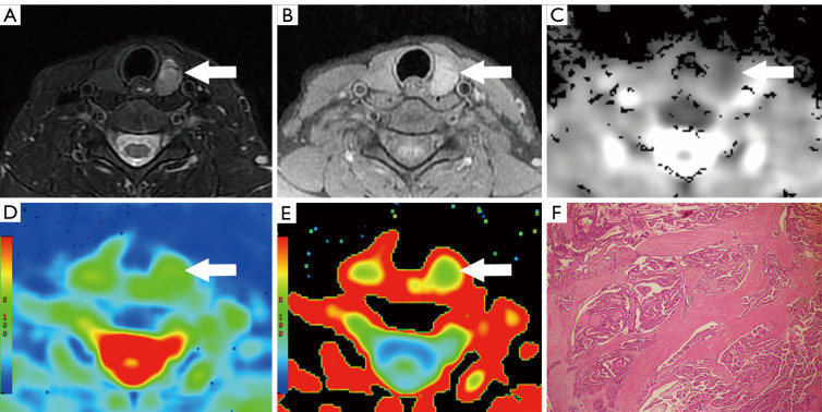
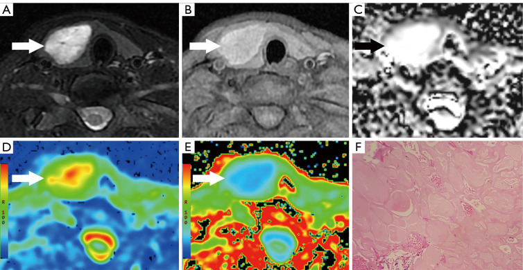
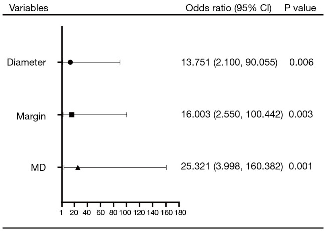
In the univariate analyses, the features that significantly indicated PTC were decreased ADC value (P<0.001), decreased MD value (P<0.001), increased MK value (P<0.001), younger age (P=0.001), female tendency (P=0.049), smaller tumor diameter (P<0.001), solid component (P<0.001), and irregular margin (P<0.001). In the multivariate analysis, decreased MD value (odds ratio =25.321; P=0.001), smaller diameter (odds ratio =13.751; P=0.006), and irregular margin (odds ratio =16.003; P=0.003) were independent risk factors for PTC. The combined predictor of MD, diameter, and margin showed an area under the receiver operating characteristic (ROC) curve of 0.996 in diagnosing PTC, with an optimal cutoff value of 0.69 (95.1% sensitivity, 100.0% specificity).
Lower MD value from DKI, smaller diameter, and irregular margin are useful predictive biomarkers for PTC.
本研究旨在探讨非增强磁共振成像(MRI)联合扩散峰度成像(DKI)在诊断甲状腺乳头状癌(PTC)中的价值。
本研究共纳入77例连续患者,共计77个甲状腺结节。其中,41个结节经组织病理学确诊为PTC,36个为良性结节。所有患者均接受甲状腺MRI检查,包括T1加权成像(T1WI)、T2加权成像(T2WI)、扩散加权成像(DWI)和DKI。所有图像均由2名放射科医生评估。测量这些结节在T1WI和T2WI上的信号强度比(SIR)、DWI的表观扩散系数(ADC)以及DKI的平均扩散率(MD)和平均峰度(MK)。还评估了这些图像上的形态学特征。采用单因素和多因素逻辑回归分析来评估这些参数作为PTC潜在预测指标的价值。
在单因素分析中,显著提示PTC的特征包括ADC值降低(P<0.001)、MD值降低(P<0.001)、MK值升高(P<0.001)、年龄较小(P=0.001)、女性倾向(P=0.049)、肿瘤直径较小(P<0.001)、实性成分(P<0.001)和边缘不规则(P<0.001)。在多因素分析中,MD值降低(比值比=25.321;P=0.001)、直径较小(比值比=13.751;P=0.006)和边缘不规则(比值比=16.003;P=0.003)是PTC的独立危险因素。MD、直径和边缘的联合预测指标在诊断PTC时的受试者操作特征(ROC)曲线下面积为0.996,最佳截断值为0.69(敏感性为95.1%,特异性为100.0%)。
DKI的较低MD值、较小直径和边缘不规则是PTC有用的预测生物标志物。