• 文献检索
  • 文档翻译
  • 深度研究
  • 学术资讯
  • Suppr Zotero 插件Zotero 插件
  • 邀请有礼
  • 套餐&价格
  • 历史记录
应用&插件
Suppr Zotero 插件Zotero 插件浏览器插件Mac 客户端Windows 客户端微信小程序
定价
高级版会员购买积分包购买API积分包
服务
文献检索文档翻译深度研究API 文档MCP 服务
关于我们
关于 Suppr公司介绍联系我们用户协议隐私条款
关注我们

Suppr 超能文献

核心技术专利:CN118964589B侵权必究
粤ICP备2023148730 号-1Suppr @ 2026

文献检索

告别复杂PubMed语法,用中文像聊天一样搜索,搜遍4000万医学文献。AI智能推荐,让科研检索更轻松。

立即免费搜索

文件翻译

保留排版,准确专业,支持PDF/Word/PPT等文件格式,支持 12+语言互译。

免费翻译文档

深度研究

AI帮你快速写综述,25分钟生成高质量综述,智能提取关键信息,辅助科研写作。

立即免费体验

理解心腔内单极电图中的复极化:重温一场长期存在的争论。

Understanding repolarization in the intracardiac unipolar electrogram: A long-lasting controversy revisited.

作者信息

Stoks Job, Bear Laura R, Vijgen Johan, Dendale Paul, Peeters Ralf, Volders Paul G A, Cluitmans Matthijs J M

机构信息

Department of Cardiology, CARIM, Maastricht University Medical Center+, Maastricht, Netherlands.

Department of Advanced Computing Sciences, Maastricht University, Maastricht, Netherlands.

出版信息

Front Physiol. 2023 Apr 7;14:1158003. doi: 10.3389/fphys.2023.1158003. eCollection 2023.

DOI:10.3389/fphys.2023.1158003
PMID:37089414
原文链接:https://pmc.ncbi.nlm.nih.gov/articles/PMC10119409/
Abstract

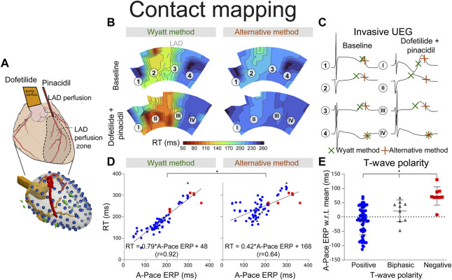
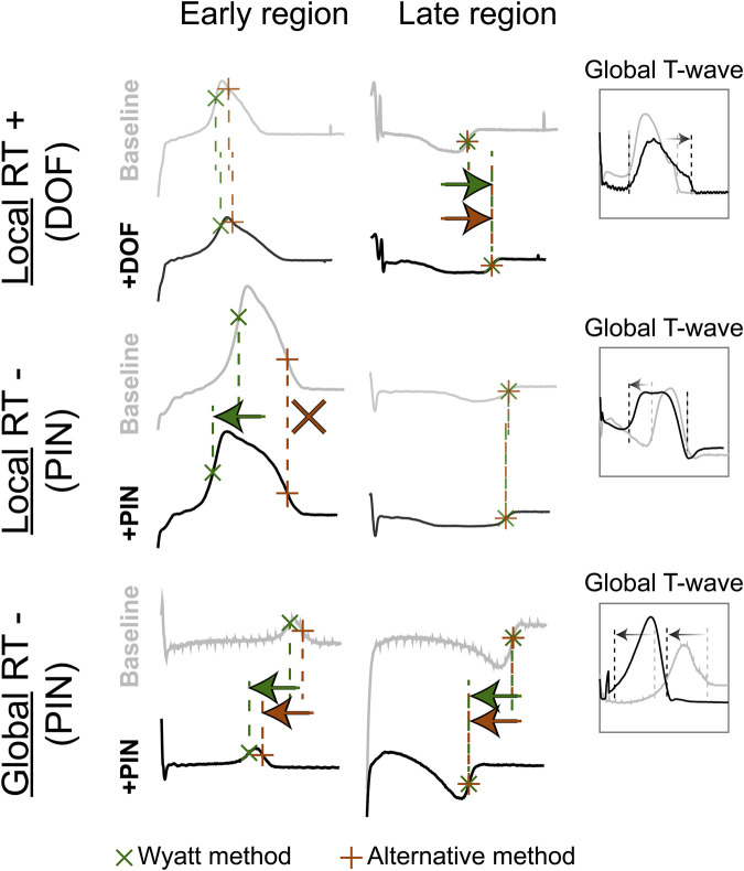
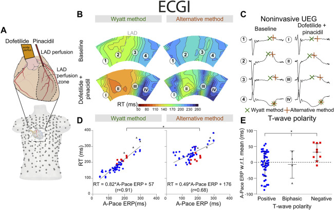
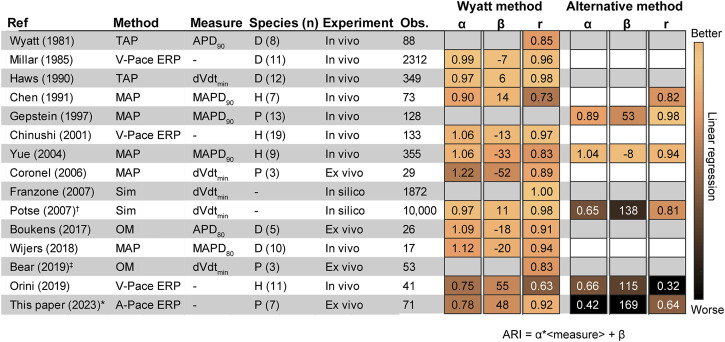
The optimal way to determine repolarization time (RT) from the intracardiac unipolar electrogram (UEG) has been a topic of debate for decades. RT is typically determined by either the Wyatt method or the "alternative method," which both consider UEG T-wave slope, but differently. To determine the optimal method to measure RT on the UEG. Seven pig hearts surrounded by an epicardial sock with 100 electrodes were Langendorff-perfused with selective cannulation of the left anterior descending (LAD) coronary artery and submersed in a torso-shaped tank containing 256 electrodes on the torso surface. Repolarization was prolonged in the non-LAD-regions by infusing dofetilide and shortened in the LAD-region using pinacidil. RT was determined by the Wyatt (t) and alternative (t) methods, in both invasive (recorded with epicardial electrodes) and in non-invasive UEGs (reconstructed with electrocardiographic imaging). t and t were compared to local effective refractory period (ERP). With contact mapping, mean absolute error (MAE) of t and t vs. ERP were 21 ms and 71 ms, respectively. Positive T-waves typically had an earlier ERP than negative T-waves, in line with theory. t -but not t-shortened by local infusion of pinacidil. Similar results were found for the non-invasive UEGs (MAE of t and t vs. ERP were 30 ms and 92 ms, respectively). The Wyatt method is the most accurate to determine RT from (non) invasive UEGs, based on novel and historical analyses. Using it to determine RT could unify and facilitate repolarization assessment and amplify its role in cardiac electrophysiology.

摘要

几十年来,从心腔内单极电图(UEG)确定复极时间(RT)的最佳方法一直是一个争论的话题。RT通常通过怀亚特方法或“替代方法”来确定,这两种方法都考虑UEG T波斜率,但方式不同。为了确定测量UEG上RT的最佳方法,对七个被带有100个电极的心外膜套包围的猪心脏进行Langendorff灌注,选择性插管左前降支(LAD)冠状动脉,并将其浸入躯干表面有256个电极的躯干形水箱中。通过输注多非利特使非LAD区域的复极延长,使用吡那地尔使LAD区域的复极缩短。RT通过怀亚特(t)方法和替代(t)方法在有创(用心外膜电极记录)和无创UEG(用心电图成像重建)中确定。将t和t与局部有效不应期(ERP)进行比较。通过接触式标测,t和t与ERP的平均绝对误差(MAE)分别为21毫秒和71毫秒。与理论一致,正向T波的ERP通常比负向T波更早。局部输注吡那地尔使t缩短,但t未缩短。无创UEG也得到了类似的结果(t和t与ERP的MAE分别为30毫秒和92毫秒)。基于新的和历史分析,怀亚特方法是从(非)侵入性UEG确定RT最准确的方法。使用它来确定RT可以统一并促进复极评估,并扩大其在心脏电生理学中的作用。

https://cdn.ncbi.nlm.nih.gov/pmc/blobs/fd57/10119409/d9f44a6d5289/fphys-14-1158003-g007.jpg
https://cdn.ncbi.nlm.nih.gov/pmc/blobs/fd57/10119409/c4f4240703e6/fphys-14-1158003-g001.jpg
https://cdn.ncbi.nlm.nih.gov/pmc/blobs/fd57/10119409/b3fce6e8206c/fphys-14-1158003-g002.jpg
https://cdn.ncbi.nlm.nih.gov/pmc/blobs/fd57/10119409/514e8db4a2c9/fphys-14-1158003-g003.jpg
https://cdn.ncbi.nlm.nih.gov/pmc/blobs/fd57/10119409/80f2fc1ff336/fphys-14-1158003-g004.jpg
https://cdn.ncbi.nlm.nih.gov/pmc/blobs/fd57/10119409/4cdada4004b6/fphys-14-1158003-g005.jpg
https://cdn.ncbi.nlm.nih.gov/pmc/blobs/fd57/10119409/5328278fa77a/fphys-14-1158003-g006.jpg
https://cdn.ncbi.nlm.nih.gov/pmc/blobs/fd57/10119409/d9f44a6d5289/fphys-14-1158003-g007.jpg

相似文献

1
Understanding repolarization in the intracardiac unipolar electrogram: A long-lasting controversy revisited.理解心腔内单极电图中的复极化:重温一场长期存在的争论。
Front Physiol. 2023 Apr 7;14:1158003. doi: 10.3389/fphys.2023.1158003. eCollection 2023.
2
Optical Imaging of Ventricular Action Potentials in a Torso Tank: A New Platform for Non-Invasive Electrocardiographic Imaging Validation.人体模型水槽中的心室外膜动作电位光学成像:用于无创心电图成像验证的新平台
Front Physiol. 2019 Feb 26;10:146. doi: 10.3389/fphys.2019.00146. eCollection 2019.
3
In vivo human sock-mapping validation of a simple model that explains unipolar electrogram morphology in relation to conduction-repolarization dynamics.体内人体袜子映射验证了一个简单的模型,该模型解释了单极电图形态与传导复极动力学的关系。
J Cardiovasc Electrophysiol. 2018 Jul;29(7):990-997. doi: 10.1111/jce.13606. Epub 2018 May 3.
4
Electrocardiographic Imaging of Repolarization Abnormalities.心电图复极异常的影像学。
J Am Heart Assoc. 2021 May 4;10(9):e020153. doi: 10.1161/JAHA.120.020153. Epub 2021 Apr 21.
5
Validation of Noninvasive Epicardial and Endocardial Repolarization Mapping.无创性心外膜和心内膜复极标测的验证
Front Physiol. 2021 Oct 22;12:737609. doi: 10.3389/fphys.2021.737609. eCollection 2021.
6
Advantages and pitfalls of noninvasive electrocardiographic imaging.无创性心电图成像的优缺点。
J Electrocardiol. 2019 Nov-Dec;57S:S15-S20. doi: 10.1016/j.jelectrocard.2019.08.007. Epub 2019 Aug 9.
7
Body Surface Mapping of Ventricular Repolarization Heterogeneity: An Multiparameter Study.心室复极异质性的体表标测:一项多参数研究。
Front Physiol. 2020 Aug 13;11:933. doi: 10.3389/fphys.2020.00933. eCollection 2020.
8
Steep repolarization time gradients in pig hearts cause distinct changes in composite electrocardiographic T-wave parameters.猪心复极时间梯度陡峭导致复合心电图 T 波参数发生明显变化。
Ann Noninvasive Electrocardiol. 2022 Nov;27(6):e12994. doi: 10.1111/anec.12994. Epub 2022 Aug 19.
9
Correlation between the effective refractory period and activation-recovery interval calculated from the intracardiac unipolar electrogram of humans with and without dl-sotalol treatment.接受与未接受dl-索他洛尔治疗的人类心腔内单极电图所计算出的有效不应期与激活-恢复间期之间的相关性。
Jpn Circ J. 2001 Aug;65(8):702-6. doi: 10.1253/jcj.65.702.
10
Dispersion of repolarization and arrhythmogenesis.复极离散与心律失常发生机制
Heart Rhythm. 2009 Apr;6(4):537-43. doi: 10.1016/j.hrthm.2009.01.013. Epub 2009 Jan 18.

引用本文的文献

1
The apicobasal dispersion of ventricular repolarization in humans is associated with age and affects arrhythmia vulnerability.人类心室复极的顶-基底部离散与年龄相关,并影响心律失常易感性。
J Physiol. 2025 May 28. doi: 10.1113/JP288356.
2
Variant patterns of electrical activation and recovery in normal human hearts revealed by noninvasive electrocardiographic imaging.非侵入性心电图成像技术揭示正常人心电活动和复极的变异模式。
Europace. 2024 Jul 2;26(7). doi: 10.1093/europace/euae172.
3
The Dutch Idiopathic Ventricular Fibrillation Registry: progress report on the quest to identify the unidentifiable.

本文引用的文献

1
Spatiotemporal approximation of cardiac activation and recovery isochrones.心脏激活和恢复等时线的时空近似。
J Electrocardiol. 2022 Mar-Apr;71:1-9. doi: 10.1016/j.jelectrocard.2021.12.007. Epub 2021 Dec 28.
2
Noninvasive detection of spatiotemporal activation-repolarization interactions that prime idiopathic ventricular fibrillation.无创检测致特发性心室颤动的时空激活-复极相互作用。
Sci Transl Med. 2021 Nov 17;13(620):eabi9317. doi: 10.1126/scitranslmed.abi9317.
3
Dynamic spatial dispersion of repolarization is present in regions critical for ischemic ventricular tachycardia ablation.
荷兰特发性心室颤动注册研究:识别难以识别者的进展报告
Neth Heart J. 2024 Jun;32(6):238-244. doi: 10.1007/s12471-024-01870-y. Epub 2024 Apr 23.
复极的动态空间离散存在于缺血性室性心动过速消融关键区域。
Heart Rhythm O2. 2021 May 11;2(3):280-289. doi: 10.1016/j.hroo.2021.05.003. eCollection 2021 Jun.
4
A novel method to correct repolarization time estimation from unipolar electrograms distorted by standard filtering.
Med Image Anal. 2021 Aug;72:102075. doi: 10.1016/j.media.2021.102075. Epub 2021 Apr 18.
5
Electrocardiographic Imaging of Repolarization Abnormalities.心电图复极异常的影像学。
J Am Heart Assoc. 2021 May 4;10(9):e020153. doi: 10.1161/JAHA.120.020153. Epub 2021 Apr 21.
6
Further Evidence on How to Measure Local Repolarization Time Using Intracardiac Unipolar Electrograms in the Intact Human Heart.关于如何在完整人体心脏中使用心腔内单极电图测量局部复极时间的进一步证据。
Circ Arrhythm Electrophysiol. 2019 Nov;12(11):e007733. doi: 10.1161/CIRCEP.119.007733. Epub 2019 Nov 8.
7
Advantages and pitfalls of noninvasive electrocardiographic imaging.无创性心电图成像的优缺点。
J Electrocardiol. 2019 Nov-Dec;57S:S15-S20. doi: 10.1016/j.jelectrocard.2019.08.007. Epub 2019 Aug 9.
8
Optical Imaging of Ventricular Action Potentials in a Torso Tank: A New Platform for Non-Invasive Electrocardiographic Imaging Validation.人体模型水槽中的心室外膜动作电位光学成像:用于无创心电图成像验证的新平台
Front Physiol. 2019 Feb 26;10:146. doi: 10.3389/fphys.2019.00146. eCollection 2019.
9
In Vivo Validation of Electrocardiographic Imaging.体内心电图成像的验证。
JACC Clin Electrophysiol. 2017 Mar;3(3):232-242. doi: 10.1016/j.jacep.2016.11.012. Epub 2017 Feb 1.
10
In vivo human sock-mapping validation of a simple model that explains unipolar electrogram morphology in relation to conduction-repolarization dynamics.体内人体袜子映射验证了一个简单的模型,该模型解释了单极电图形态与传导复极动力学的关系。
J Cardiovasc Electrophysiol. 2018 Jul;29(7):990-997. doi: 10.1111/jce.13606. Epub 2018 May 3.