IHU-LIRYCFondation Bordeaux Université Pessac France.
CRCTB U1045 Université de Bordeaux Bordeaux France.
J Am Heart Assoc. 2021 May 4;10(9):e020153. doi: 10.1161/JAHA.120.020153. Epub 2021 Apr 21.
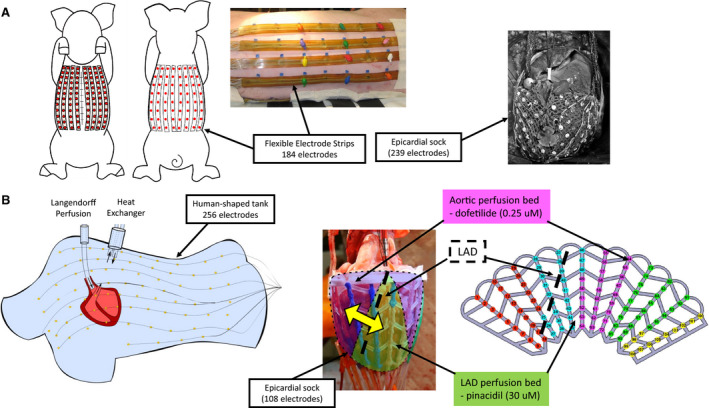
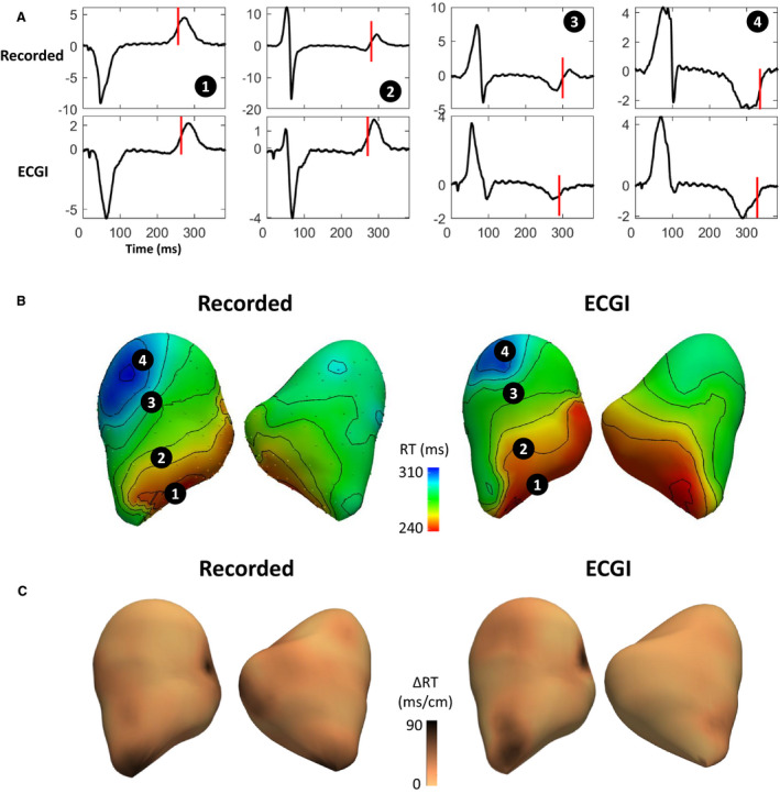
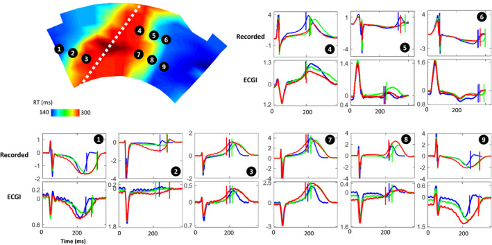
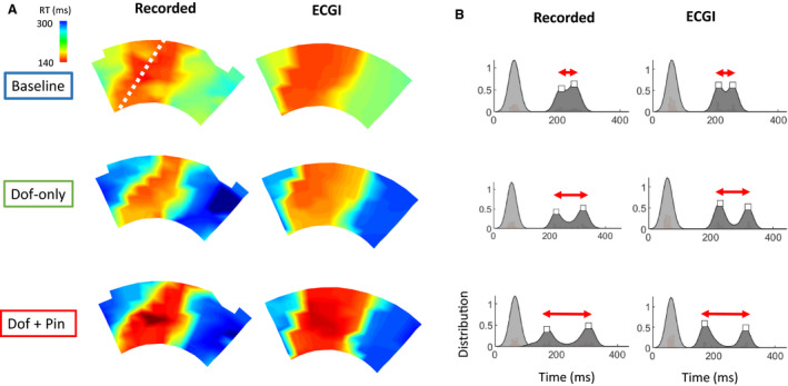
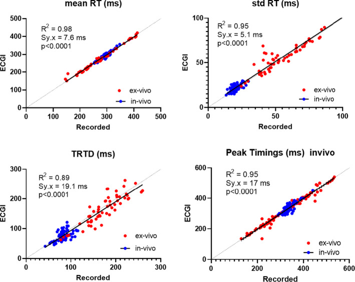
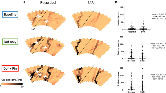
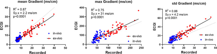
Background Dispersion and gradients in repolarization have been associated with life-threatening arrhythmias, but are difficult to quantify precisely from surface electrocardiography. The objective of this study was to evaluate electrocardiographic imaging (ECGI) to noninvasively detect repolarization-based abnormalities. Methods and Results Ex vivo data were obtained from Langendorff-perfused pig hearts (n=8) and a human donor heart. Unipolar electrograms were recorded simultaneously during sinus rhythm from an epicardial sock and the torso-shaped tank within which the heart was suspended. Regional repolarization heterogeneities were introduced through perfusion of dofetilide and pinacidil into separate perfusion beds. In vivo data included torso and epicardial potentials recorded simultaneously in anesthetized, closed-chest pigs (n=5), during sinus rhythm, and ventricular pacing. For both data sets, ECGI accurately reconstructed T-wave electrogram morphologies when compared with those recorded by the sock (ex vivo: correlation coefficient, 0.85 [0.52-0.96], in vivo: correlation coefficient, 0.86 [0.52-0.96]) and repolarization time maps (ex-vivo: correlation coefficient, 0.73 [0.63-0.83], in vivo: correlation coefficient, 0.76 [0.67-0.82]). ECGI-reconstructed repolarization time distributions were strongly correlated to those measured by the sock (both data sets, ≥0.92). Although the position of the gradient was slightly shifted by 8.3 (0-13.9) mm, the mean, max, and SD between ECGI and recorded gradient values were highly correlated (=0.87, 0.75, and 0.86 respectively). There was no significant difference in ECGI accuracy between ex vivo and in vivo data. Conclusions ECGI reliably and accurately maps potentially critical repolarization abnormalities. This noninvasive approach allows imaging and quantifying individual parameters of abnormal repolarization-based substrates in patients with arrhythmogenesis, to improve diagnosis and risk stratification.
复极离散度和梯度与危及生命的心律失常有关,但很难通过体表心电图精确量化。本研究旨在评估心电图成像(ECGI)技术以无创方式检测复极异常。
离体数据来自 Langendorff 灌注猪心(n=8)和人类供心。窦性心律时,同时从心外膜套和悬挂心脏的体形容器内记录单极电图。通过向不同的灌注床灌注多非利特和匹那地尔来引入区域性复极异质性。在体内数据中,包括在麻醉、闭胸猪(n=5)中记录的同步心外膜和心外膜电位,窦性心律和心室起搏时。对于这两个数据集,与套(离体:相关系数 0.85 [0.52-0.96],体内:相关系数 0.86 [0.52-0.96])和复极时间图(离体:相关系数 0.73 [0.63-0.83],体内:相关系数 0.76 [0.67-0.82])相比,ECGI 准确地重建了 T 波电图形态。ECGI 重建的复极时间分布与套(两个数据集,≥0.92)测量的复极时间分布高度相关。虽然梯度的位置略微偏移了 8.3(0-13.9)mm,但 ECGI 与记录梯度值之间的平均值、最大值和标准差高度相关(=0.87、0.75 和 0.86 分别)。离体和体内数据之间的 ECGI 准确性没有显著差异。
ECGI 可靠且准确地绘制潜在的临界复极异常图。这种非侵入性方法可以对心律失常发生患者的异常复极基础进行成像和定量个体参数,以改善诊断和风险分层。