Department of Mechanical Engineering, University College London, London, United Kingdom.
Department of Cardiac Electrophysiology, The Barts Heart Center, St Bartholomew's Hospital, London, United Kingdom.
J Cardiovasc Electrophysiol. 2018 Jul;29(7):990-997. doi: 10.1111/jce.13606. Epub 2018 May 3.
The unipolar electrogram (UEG) provides local measures of cardiac activation and repolarization and is an important translational link between patient and laboratory. A simple theoretical model of the UEG was previously proposed and tested in silico.
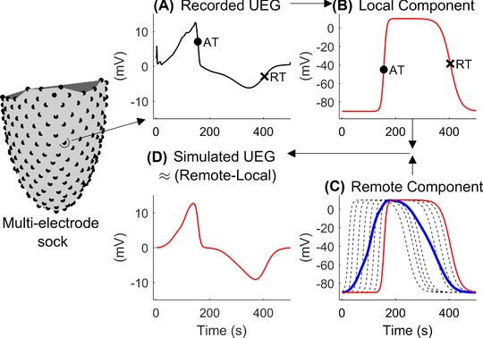
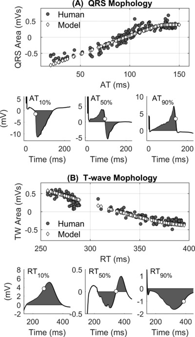
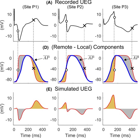
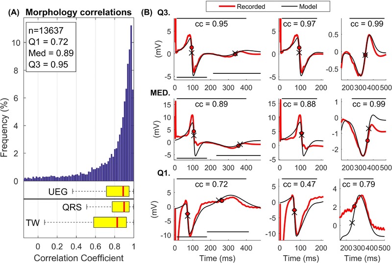
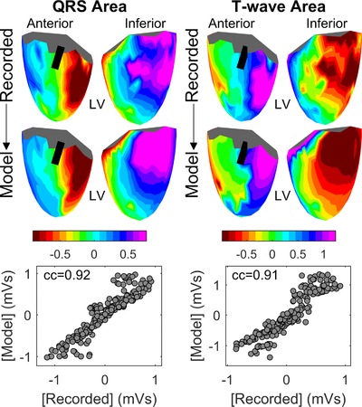
The aim of this study was to use epicardial sock-mapping data to validate the simple model's predictions of unipolar electrogram morphology in the in vivo human heart. The simple model conceptualizes the UEG as the difference between a local cardiac action potential and a position-independent component representing remote activity, which is defined as the average of all action potentials. UEGs were recorded in 18 patients using a multielectrode sock containing 240 electrodes and activation (AT) and repolarization time (RT) were measured using standard definitions. For each cardiac site, a simulated local action potential was generated by adjusting a stylized action potential to fit AT and RT measured in vivo. The correlation coefficient (cc) measuring the morphological similarity between 13,637 recorded and simulated UEGs was cc = 0.89 (0.72-0.95), median (Q -Q ), for the entire UEG, cc = 0.90 (0.76-0.95) for QRS complexes, and cc = 0.83 (0.58-0.92) for T-waves. QRS and T-wave areas from recorded and simulated UEGs showed cc> 0.89 and cc> 0.84, respectively, indicating good agreement between voltage isochrones maps. Simulated UEGs accurately reproduced the interaction between AT and QRS morphology and between RT and T-wave morphology observed in vivo.
Human in vivo whole heart data support the validity of the simple model, which provides a framework for improving the understanding of the UEG and its clinical utility.
单极电图(UEG)提供了心脏激活和复极的局部测量值,是将患者与实验室联系起来的重要转化环节。之前已经提出并在计算机上测试了一种简单的 UEG 理论模型。
本研究的目的是使用心外膜套状标测数据来验证简单模型对体内人心率 UEG 形态的预测。该简单模型将 UEG 概念化为局部心动作电位与代表远程活动的位置独立分量之间的差异,该分量定义为所有动作电位的平均值。使用包含 240 个电极的多电极套记录 18 名患者的 UEG,并使用标准定义测量激活(AT)和复极时间(RT)。对于每个心脏部位,通过调整样式化动作电位以拟合体内测量的 AT 和 RT 来生成模拟的局部动作电位。测量 13637 个记录和模拟 UEG 之间形态相似性的相关系数(cc)为整个 UEG 的 cc=0.89(0.72-0.95),中位数(Q-Q),对于 QRS 复合体,cc=0.90(0.76-0.95),对于 T 波,cc=0.83(0.58-0.92)。记录和模拟 UEG 的 QRS 和 T 波区域的 cc 值均>0.89 和 cc 值>0.84,表明电压等时线图之间具有良好的一致性。模拟的 UEG 准确地再现了体内观察到的 AT 与 QRS 形态之间以及 RT 与 T 波形态之间的相互作用。
人体整体心脏数据支持简单模型的有效性,该模型为理解 UEG 及其临床应用提供了框架。