Diao Kaiyue, Liang Hong-Qing, Yin Hong-Kun, Yuan Ming-Jing, Gu Min, Yu Peng-Xin, He Sen, Sun Jiayu, Song Bin, Li Kang, He Yong
Department of Radiology, West China Hospital of Sichuan University, Chengdu, Sichuan, China.
Department of Radiology, First Affiliated Hospital to Army Medical University (Third Military Medical University Southwest Hospital), Chongqing, China.
Insights Imaging. 2023 Apr 24;14(1):70. doi: 10.1186/s13244-023-01401-0.
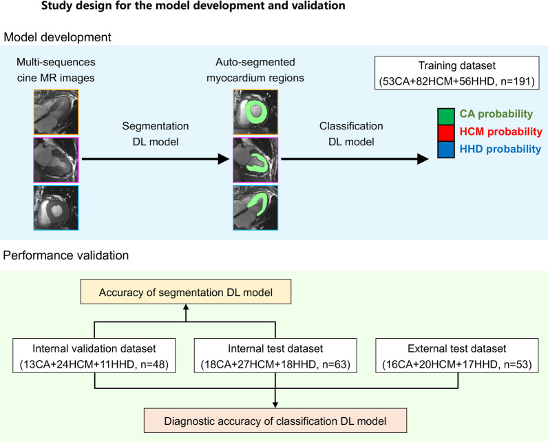
To develop a fully automatic framework for the diagnosis of cause for left ventricular hypertrophy (LVH) via cardiac cine images.
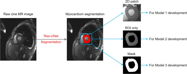
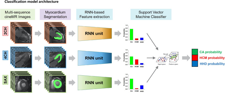
A total of 302 LVH patients with cine MRI images were recruited as the primary cohort. Another 53 LVH patients prospectively collected or from multi-centers were used as the external test dataset. Different models based on the cardiac regions (Model 1), segmented ventricle (Model 2) and ventricle mask (Model 3) were constructed. The diagnostic performance was accessed by the confusion matrix with respect to overall accuracy. The capability of the predictive models for binary classification of cardiac amyloidosis (CA), hypertrophic cardiomyopathy (HCM) or hypertensive heart disease (HHD) were also evaluated. Additionally, the diagnostic performance of best Model was compared with that of 7 radiologists/cardiologists.
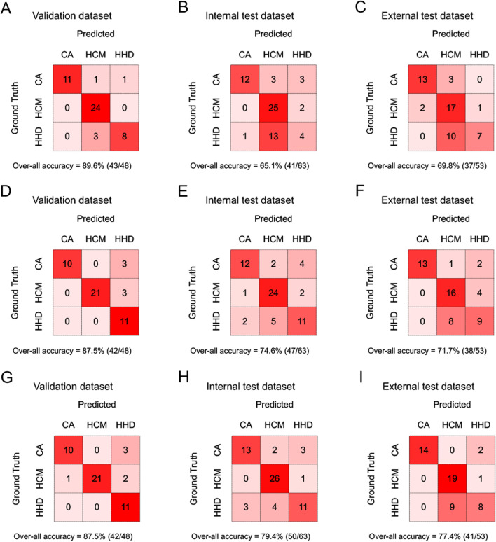
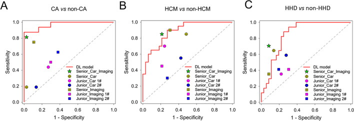
Model 3 showed the best performance with an overall classification accuracy up to 77.4% in the external test datasets. On the subtasks for identifying CA, HCM or HHD only, Model 3 also achieved the best performance with AUCs yielding 0.895-0.980, 0.879-0.984 and 0.848-0.983 in the validation, internal test and external test datasets, respectively. The deep learning model showed non-inferior diagnostic capability to the cardiovascular imaging expert and outperformed other radiologists/cardiologists.
The combined model based on the mask of left ventricular segmented from multi-sequences cine MR images shows favorable and robust performance in diagnosing the cause of left ventricular hypertrophy, which could be served as a noninvasive tool and help clinical decision.
开发一种通过心脏电影图像诊断左心室肥厚(LVH)病因的全自动框架。
招募了302例有电影MRI图像的LVH患者作为主要队列。另外53例前瞻性收集的或来自多中心的LVH患者用作外部测试数据集。构建了基于心脏区域的不同模型(模型1)、分割心室模型(模型2)和心室掩码模型(模型3)。通过混淆矩阵评估总体准确性的诊断性能。还评估了预测模型对心脏淀粉样变性(CA)、肥厚型心肌病(HCM)或高血压性心脏病(HHD)进行二元分类的能力。此外,将最佳模型的诊断性能与7位放射科医生/心脏病专家的诊断性能进行了比较。
模型3表现最佳,在外部测试数据集中总体分类准确率高达77.4%。仅在识别CA、HCM或HHD的子任务上,模型3也表现最佳,在验证数据集、内部测试数据集和外部测试数据集中的AUC分别为0.895 - 0.980、0.879 - 0.984和0.848 - 0.983。深度学习模型显示出与心血管影像专家相当的诊断能力,并且优于其他放射科医生/心脏病专家。
基于多序列电影MR图像分割的左心室掩码的组合模型在诊断左心室肥厚病因方面表现出良好且稳健的性能,可作为一种无创工具并有助于临床决策。