• 文献检索
  • 文档翻译
  • 深度研究
  • 学术资讯
  • Suppr Zotero 插件Zotero 插件
  • 邀请有礼
  • 套餐&价格
  • 历史记录
应用&插件
Suppr Zotero 插件Zotero 插件浏览器插件Mac 客户端Windows 客户端微信小程序
定价
高级版会员购买积分包购买API积分包
服务
文献检索文档翻译深度研究API 文档MCP 服务
关于我们
关于 Suppr公司介绍联系我们用户协议隐私条款
关注我们

Suppr 超能文献

核心技术专利:CN118964589B侵权必究
粤ICP备2023148730 号-1Suppr @ 2026

文献检索

告别复杂PubMed语法,用中文像聊天一样搜索,搜遍4000万医学文献。AI智能推荐,让科研检索更轻松。

立即免费搜索

文件翻译

保留排版,准确专业,支持PDF/Word/PPT等文件格式,支持 12+语言互译。

免费翻译文档

深度研究

AI帮你快速写综述,25分钟生成高质量综述,智能提取关键信息,辅助科研写作。

立即免费体验

Svep1 是 Tie1 的结合配体,以独立于 Vegfc 的方式影响面部淋巴管发育的特定方面。

Svep1 is a binding ligand of Tie1 and affects specific aspects of facial lymphatic development in a Vegfc-independent manner.

机构信息

Institute of Cardiovascular Organogenesis and Regeneration, Faculty of Medicine, WWU Münster, Münster, Germany.

Münster Imaging Network, Cells in Motion Interfaculty Centre, Faculty of Biology, WWU Münster, Münster, Germany.

出版信息

Elife. 2023 Apr 25;12:e82969. doi: 10.7554/eLife.82969.

DOI:10.7554/eLife.82969
PMID:37097004
原文链接:https://pmc.ncbi.nlm.nih.gov/articles/PMC10129328/
Abstract

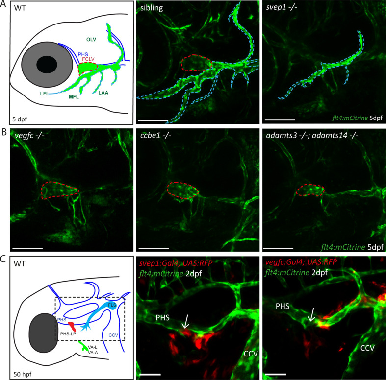
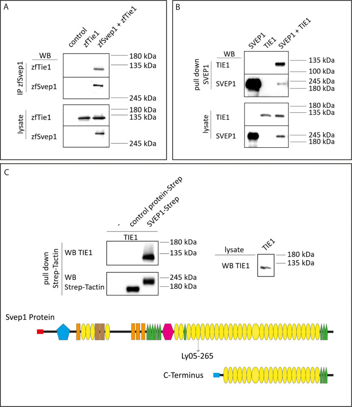
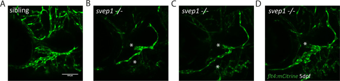
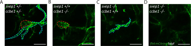
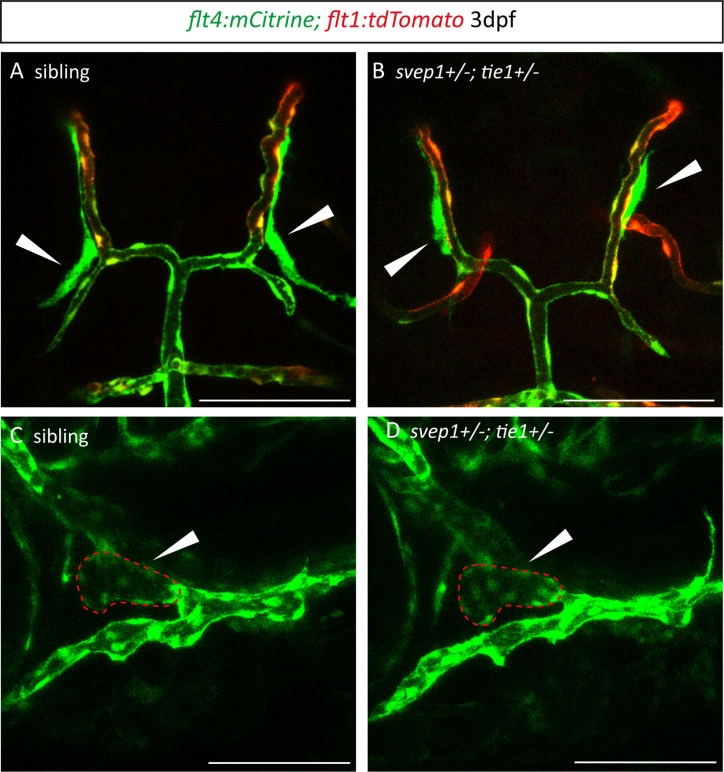
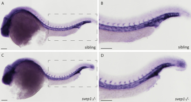
Multiple factors are required to form functional lymphatic vessels. Here, we uncover an essential role for the secreted protein Svep1 and the transmembrane receptor Tie1 during the development of subpopulations of the zebrafish facial lymphatic network. This specific aspect of the facial network forms independently of Vascular endothelial growth factor C (Vegfc) signalling, which otherwise is the most prominent signalling axis in all other lymphatic beds. Additionally, we find that multiple specific and newly uncovered phenotypic hallmarks of mutants are also present in , but not in or mutants. These phenotypes are observed in the lymphatic vasculature of both head and trunk, as well as in the development of the dorsal longitudinal anastomotic vessel under reduced flow conditions. Therefore, our study demonstrates an important function for Tie1 signalling during lymphangiogenesis as well as blood vessel development in zebrafish. Furthermore, we show genetic interaction between and in vivo, during early steps of lymphangiogenesis, and demonstrate that zebrafish as well as human Svep1/SVEP1 protein bind to the respective Tie1/TIE1 receptors in vitro. Since compound heterozygous mutations for and have recently been reported in human glaucoma patients, our data have clinical relevance in demonstrating a role for SVEP1 in TIE signalling in an in vivo setting.

摘要

多种因素是形成功能性淋巴管所必需的。在这里,我们揭示了分泌蛋白 Svep1 和跨膜受体 Tie1 在斑马鱼面部淋巴管网络亚群发育过程中的重要作用。面部网络的这一特定方面的形成独立于血管内皮生长因子 C(Vegfc)信号,而 Vegfc 信号是所有其他淋巴管床中最突出的信号轴。此外,我们发现, 突变体的多个特定和新发现的表型特征也存在于 突变体中,但不存在于 或 突变体中。这些表型在头部和躯干的淋巴管血管以及在减少血流条件下背侧纵向吻合血管的发育中都可以观察到。因此,我们的研究表明 Tie1 信号在斑马鱼淋巴管生成以及血管发育过程中具有重要功能。此外,我们还在体内早期的淋巴管生成过程中显示了 和 之间的遗传相互作用,并证明了斑马鱼和人类 Svep1/SVEP1 蛋白在体外与相应的 Tie1/TIE1 受体结合。由于 和 的复合杂合突变最近在人类青光眼患者中被报道,我们的数据在证明 SVEP1 在体内 TIE 信号中的作用方面具有临床相关性。

https://cdn.ncbi.nlm.nih.gov/pmc/blobs/145e/10129328/d5e333b8bd0e/elife-82969-fig8.jpg
https://cdn.ncbi.nlm.nih.gov/pmc/blobs/145e/10129328/6a46f5e3b963/elife-82969-fig1.jpg
https://cdn.ncbi.nlm.nih.gov/pmc/blobs/145e/10129328/2292f82b2570/elife-82969-fig1-figsupp1.jpg
https://cdn.ncbi.nlm.nih.gov/pmc/blobs/145e/10129328/7cfedb0e5266/elife-82969-fig1-figsupp2.jpg
https://cdn.ncbi.nlm.nih.gov/pmc/blobs/145e/10129328/19fd99be5b6b/elife-82969-fig1-figsupp3.jpg
https://cdn.ncbi.nlm.nih.gov/pmc/blobs/145e/10129328/4d75cdc8b3d8/elife-82969-fig2.jpg
https://cdn.ncbi.nlm.nih.gov/pmc/blobs/145e/10129328/4d119af2d6a1/elife-82969-fig3.jpg
https://cdn.ncbi.nlm.nih.gov/pmc/blobs/145e/10129328/7d4dbc6cdcb1/elife-82969-fig3-figsupp1.jpg
https://cdn.ncbi.nlm.nih.gov/pmc/blobs/145e/10129328/4fea877fe866/elife-82969-fig4.jpg
https://cdn.ncbi.nlm.nih.gov/pmc/blobs/145e/10129328/dedda778fc70/elife-82969-fig4-figsupp1.jpg
https://cdn.ncbi.nlm.nih.gov/pmc/blobs/145e/10129328/77a9d2ab5339/elife-82969-fig4-figsupp2.jpg
https://cdn.ncbi.nlm.nih.gov/pmc/blobs/145e/10129328/f9294f233901/elife-82969-fig5.jpg
https://cdn.ncbi.nlm.nih.gov/pmc/blobs/145e/10129328/30ca4bb8495b/elife-82969-fig5-figsupp1.jpg
https://cdn.ncbi.nlm.nih.gov/pmc/blobs/145e/10129328/5ad192ce0e43/elife-82969-fig5-figsupp2.jpg
https://cdn.ncbi.nlm.nih.gov/pmc/blobs/145e/10129328/800981274085/elife-82969-fig5-figsupp3.jpg
https://cdn.ncbi.nlm.nih.gov/pmc/blobs/145e/10129328/d9f30d6bdf04/elife-82969-fig6.jpg
https://cdn.ncbi.nlm.nih.gov/pmc/blobs/145e/10129328/a92bfc7c68af/elife-82969-fig7.jpg
https://cdn.ncbi.nlm.nih.gov/pmc/blobs/145e/10129328/477f26a3be52/elife-82969-fig7-figsupp1.jpg
https://cdn.ncbi.nlm.nih.gov/pmc/blobs/145e/10129328/139b46204313/elife-82969-fig7-figsupp2.jpg
https://cdn.ncbi.nlm.nih.gov/pmc/blobs/145e/10129328/d5e333b8bd0e/elife-82969-fig8.jpg

相似文献

1
Svep1 is a binding ligand of Tie1 and affects specific aspects of facial lymphatic development in a Vegfc-independent manner.Svep1 是 Tie1 的结合配体,以独立于 Vegfc 的方式影响面部淋巴管发育的特定方面。
Elife. 2023 Apr 25;12:e82969. doi: 10.7554/eLife.82969.
2
Angpt1 binding to Tie1 regulates the signaling required for lymphatic vessel development in zebrafish.血管生成素 1(Angpt1)与血管生成素 1 受体(Tie1)结合,调节斑马鱼淋巴管发育所需的信号通路。
Development. 2024 May 15;151(10). doi: 10.1242/dev.202269. Epub 2024 May 28.
3
Polydom Is an Extracellular Matrix Protein Involved in Lymphatic Vessel Remodeling.多域蛋白是一种参与淋巴管重塑的细胞外基质蛋白。
Circ Res. 2017 Apr 14;120(8):1276-1288. doi: 10.1161/CIRCRESAHA.116.308825. Epub 2017 Feb 8.
4
An Evolutionarily Conserved Role for Polydom/Svep1 During Lymphatic Vessel Formation.多结构域蛋白/ SVEp1在淋巴管形成过程中具有进化保守作用。
Circ Res. 2017 Apr 14;120(8):1263-1275. doi: 10.1161/CIRCRESAHA.116.308813. Epub 2017 Feb 8.
5
Genetic dissection of tie pathway in mouse lymphatic maturation and valve development.在小鼠淋巴管成熟和瓣膜发育过程中对 Tie 通路进行遗传剖析。
Arterioscler Thromb Vasc Biol. 2014 Jun;34(6):1221-30. doi: 10.1161/ATVBAHA.113.302923. Epub 2014 Apr 24.
6
Specific fibroblast subpopulations and neuronal structures provide local sources of Vegfc-processing components during zebrafish lymphangiogenesis.在斑马鱼淋巴管生成过程中,特定的成纤维细胞亚群和神经元结构为 Vegfc 加工成分提供了局部来源。
Nat Commun. 2020 Jun 1;11(1):2724. doi: 10.1038/s41467-020-16552-7.
7
The RNA helicase Ddx21 controls Vegfc-driven developmental lymphangiogenesis by balancing endothelial cell ribosome biogenesis and p53 function.RNA 解旋酶 Ddx21 通过平衡内皮细胞核糖体生物发生和 p53 功能来控制 Vegfc 驱动的发育性淋巴管生成。
Nat Cell Biol. 2021 Nov;23(11):1136-1147. doi: 10.1038/s41556-021-00784-w. Epub 2021 Nov 8.
8
Tie1 regulates the Tie2 agonistic role of angiopoietin-2 in human lymphatic endothelial cells.Tie1 调节血管生成素-2 对人淋巴管内皮细胞的 Tie2 激动作用。
Biochem Biophys Res Commun. 2012 Mar 9;419(2):281-6. doi: 10.1016/j.bbrc.2012.02.009. Epub 2012 Feb 10.
9
Polydom/SVEP1 binds to Tie1 and promotes migration of lymphatic endothelial cells.多域/SVEP1 与 Tie1 结合,促进淋巴管内皮细胞迁移。
J Cell Biol. 2023 Sep 4;222(9). doi: 10.1083/jcb.202208047. Epub 2023 Jun 20.
10
Vegfd can compensate for loss of Vegfc in zebrafish facial lymphatic sprouting.Vegfd 可补偿斑马鱼面部淋巴管发芽过程中 Vegfc 的缺失。
Development. 2014 Jul;141(13):2680-90. doi: 10.1242/dev.106591. Epub 2014 Jun 5.

引用本文的文献

1
SVEP1 Upregulation Is a Prominent Molecular Response to Dietary Sodium Loading and Correlates with Inverse Salt Sensitivity.SVEP1上调是对饮食中钠负荷的显著分子反应,且与盐敏感性呈负相关。
medRxiv. 2025 Aug 14:2025.08.12.25333544. doi: 10.1101/2025.08.12.25333544.
2
TIE1-dependent lymphatic vascular remodeling is mediated by its second tyrosine kinase domain.TIE1 依赖性淋巴管重塑由其第二个酪氨酸激酶结构域介导。
Development. 2025 Jul 1;152(13). doi: 10.1242/dev.204469. Epub 2025 Jun 27.
3
Idiopathic multicentric Castleman disease - TAFRO results in high levels of mTOR activator SVEP1, tissue factor, and endotheliopathy.

本文引用的文献

1
Lymphangiogenesis requires Ang2/Tie/PI3K signaling for VEGFR3 cell-surface expression.淋巴管生成需要 Ang2/Tie/PI3K 信号通路来促进 VEGFR3 的细胞表面表达。
J Clin Invest. 2022 Aug 1;132(15). doi: 10.1172/JCI155478.
2
Svep1 stabilises developmental vascular anastomosis in reduced flow conditions.Svep1 稳定了低血流条件下发育性血管吻合。
Development. 2022 Mar 15;149(6). doi: 10.1242/dev.199858. Epub 2022 Mar 24.
3
Cellular crosstalk regulates the aqueous humor outflow pathway and provides new targets for glaucoma therapies.
特发性多中心Castleman病 - TAFRO综合征导致高水平的mTOR激活剂SVEP1、组织因子和内皮病变。
Blood Vessel Thromb Hemost. 2024 Jun;1(2). doi: 10.1016/j.bvth.2024.100006. Epub 2024 Apr 3.
4
Epicardial VEGFC/D signaling is essential for coronary lymphangiogenesis.心外膜VEGFC/D信号传导对于冠状淋巴管生成至关重要。
EMBO Rep. 2025 Mar 24. doi: 10.1038/s44319-025-00431-7.
5
Deep plasma and tissue proteome profiling of knockout mice reveals pathways associated with deficiency.基因敲除小鼠的深度血浆和组织蛋白质组分析揭示了与缺陷相关的途径。
J Mol Cell Cardiol Plus. 2025 Jan 10;11:100283. doi: 10.1016/j.jmccpl.2025.100283. eCollection 2025 Mar.
6
Lymphatic System Development and Function.淋巴系统发育与功能。
Curr Cardiol Rep. 2024 Nov;26(11):1209-1219. doi: 10.1007/s11886-024-02120-8. Epub 2024 Aug 22.
7
The Alzheimer's disease-linked protease BACE2 cleaves VEGFR3 and modulates its signaling.阿尔茨海默病相关蛋白酶 BACE2 切割 VEGFR3 并调节其信号转导。
J Clin Invest. 2024 Jun 18;134(16):e170550. doi: 10.1172/JCI170550.
8
Multiple cis-regulatory elements control prox1a expression in distinct lymphatic vascular beds.多个顺式调控元件控制特定淋巴管床中 prox1a 的表达。
Development. 2024 May 1;151(9). doi: 10.1242/dev.202525. Epub 2024 May 9.
9
Lymphatic vessel: origin, heterogeneity, biological functions, and therapeutic targets.淋巴管:起源、异质性、生物学功能和治疗靶点。
Signal Transduct Target Ther. 2024 Jan 3;9(1):9. doi: 10.1038/s41392-023-01723-x.
10
The emerging Janus face of SVEP1 in development and disease.SVEP1 在发育和疾病中的新兴双面性。
Trends Mol Med. 2023 Nov;29(11):939-950. doi: 10.1016/j.molmed.2023.08.002. Epub 2023 Sep 4.
细胞串扰调节房水流出通路,并为青光眼治疗提供新靶点。
Nat Commun. 2021 Oct 18;12(1):6072. doi: 10.1038/s41467-021-26346-0.
4
SVEP1 is important for morphogenesis of lymphatic system: Possible implications in lymphedema.SVEP1对淋巴系统的形态发生很重要:对淋巴水肿的潜在影响。
Lymphology. 2021;54(1):12-22.
5
Lymphatic Malformations: Genetics, Mechanisms and Therapeutic Strategies.淋巴畸形:遗传学、机制与治疗策略。
Circ Res. 2021 Jun 25;129(1):136-154. doi: 10.1161/CIRCRESAHA.121.318142. Epub 2021 Jun 24.
6
A Small Molecule Inhibitor of VE-PTP Activates Tie2 in Schlemm's Canal Increasing Outflow Facility and Reducing Intraocular Pressure.一种小分子 VE-PTP 抑制剂激活小梁网中的 Tie2,增加房水流出效率,降低眼压。
Invest Ophthalmol Vis Sci. 2020 Dec 1;61(14):12. doi: 10.1167/iovs.61.14.12.
7
SVEP1 as a Genetic Modifier of TEK-Related Primary Congenital Glaucoma.SVEP1 作为 TEK 相关原发性先天性青光眼的遗传修饰因子。
Invest Ophthalmol Vis Sci. 2020 Oct 1;61(12):6. doi: 10.1167/iovs.61.12.6.
8
Tie1 regulates zebrafish cardiac morphogenesis through Tolloid-like 1 expression.Tie1 通过 Tolloid-like 1 表达调控斑马鱼心脏形态发生。
Dev Biol. 2021 Jan 1;469:54-67. doi: 10.1016/j.ydbio.2020.09.008. Epub 2020 Sep 21.
9
Apelin signaling drives vascular endothelial cells toward a pro-angiogenic state.Apelin 信号促使血管内皮细胞向促血管生成状态发展。
Elife. 2020 Sep 21;9:e55589. doi: 10.7554/eLife.55589.
10
Tek (Tie2) is not required for cardiovascular development in zebrafish.Tek(Tie2)在斑马鱼心血管发育中不是必需的。
Development. 2020 Oct 12;147(19):dev193029. doi: 10.1242/dev.193029.