Lamichhane Bidhan, Luckett Patrick H, Dierker Donna, Yun Park Ki, Burton Harold, Olufawo Michael, Trevino Gabriel, Lee John J, Daniel Andy G S, Hacker Carl D, Marcus Daniel S, Shimony Joshua S, Leuthardt Eric C
Department of Neurological Surgery, Washington University School of Medicine, St. Louis, Missouri, USA.
Mallinckrodt Institute of Radiology, Washington University School of Medicine, St. Louis, Missouri, USA.
Neurooncol Adv. 2023 Apr 14;5(1):vdad034. doi: 10.1093/noajnl/vdad034. eCollection 2023 Jan-Dec.
Patients with glioblastoma (GBM) and high-grade glioma (HGG, World Health Organization [WHO] grade IV glioma) have a poor prognosis. Consequently, there is an unmet clinical need for accessible and noninvasively acquired predictive biomarkers of overall survival in patients. This study evaluated morphological changes in the brain separated from the tumor invasion site (ie, contralateral hemisphere). Specifically, we examined the prognostic value of widespread alterations of cortical thickness (CT) in GBM/HGG patients.
We used FreeSurfer, applied with high-resolution T1-weighted MRI, to examine CT, evaluated prior to standard treatment with surgery and chemoradiation in patients (GBM/HGG, = 162, mean age 61.3 years) and 127 healthy controls (HC; 61.9 years mean age). We then compared CT in patients to HC and studied patients' associated changes in CT as a potential biomarker of overall survival.
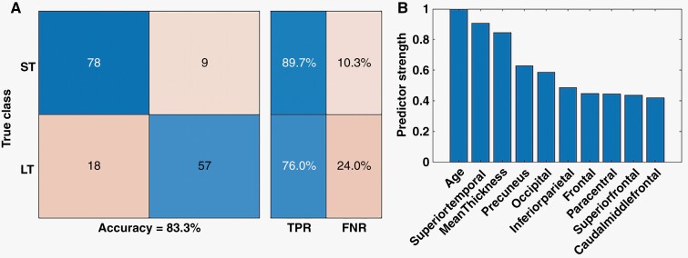
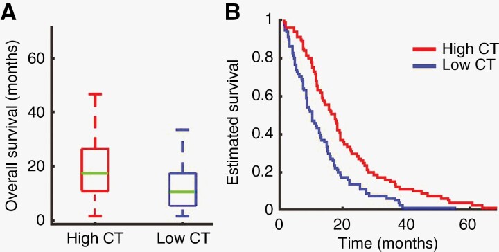
Compared to HC cases, patients had thinner gray matter in the contralesional hemisphere at the time of tumor diagnosis. patients had significant cortical thinning in parietal, temporal, and occipital lobes. Fourteen cortical parcels showed reduced CT, whereas in 5, it was thicker in patients' cases. Notably, CT in the contralesional hemisphere, various lobes, and parcels was predictive of overall survival. A machine learning classification algorithm showed that CT could differentiate short- and long-term survival patients with an accuracy of 83.3%.
These findings identify previously unnoticed structural changes in the cortex located in the hemisphere contralateral to the primary tumor mass. Observed changes in CT may have prognostic value, which could influence care and treatment planning for individual patients.
胶质母细胞瘤(GBM)和高级别胶质瘤(HGG,世界卫生组织[WHO]IV级胶质瘤)患者预后较差。因此,临床上对于可获取的、非侵入性获得的患者总生存预测生物标志物存在未满足的需求。本研究评估了与肿瘤侵袭部位分离的脑区(即对侧半球)的形态学变化。具体而言,我们研究了GBM/HGG患者广泛的皮质厚度(CT)改变的预后价值。
我们使用FreeSurfer软件,结合高分辨率T1加权磁共振成像(MRI)来检查CT,在患者(GBM/HGG,n = 162,平均年龄61.3岁)和127名健康对照者(HC;平均年龄61.9岁)接受手术及放化疗的标准治疗前进行评估。然后我们将患者的CT与HC进行比较,并研究患者CT的相关变化作为总生存的潜在生物标志物。
与HC相比,患者在肿瘤诊断时对侧半球的灰质较薄。患者在顶叶、颞叶和枕叶有明显的皮质变薄。14个皮质区域的CT降低,而在5个区域,患者的CT更厚。值得注意的是,对侧半球以及各个脑叶和区域的CT可预测总生存。一种机器学习分类算法显示,CT能够以83.3%的准确率区分短期和长期生存的患者。
这些发现揭示了位于原发肿瘤对侧半球皮质中以前未被注意到的结构变化。观察到的CT变化可能具有预后价值,这可能会影响个体患者的护理和治疗计划。