Sobue Akira, Komine Okiru, Yamanaka Koji
Department of Neuroscience and Pathobiology, Research Institute of Environmental Medicine, Nagoya University, Aichi, 464-8601, Japan.
Department of Neuroscience and Pathobiology, Nagoya University Graduate School of Medicine, Aichi, 466-8550, Japan.
Inflamm Regen. 2023 May 10;43(1):26. doi: 10.1186/s41232-023-00277-3.
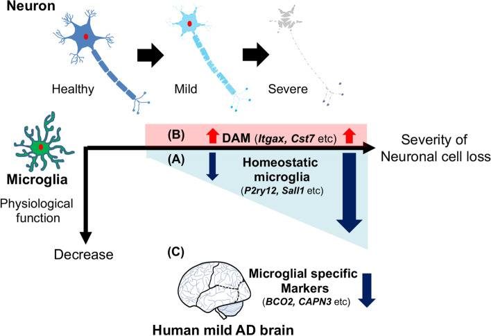
Alzheimer's disease (AD) is the most common form of dementia, pathologically characterized by senile plaques and neurofibrillary tangles (NFTs), resulting in neurodegeneration. Neuroinflammation, defined as the activation of glial cells such as microglia and astrocytes, is observed surrounding senile plaques and affected neurons in AD. Recently conducted genome-wide association studies (GWAS) indicate that a large section of identified AD risk genes are involved in immune responses and are enriched in microglia. Microglia are innate immune cells in the central nervous system (CNS), which are involved in immune surveillance and maintenance of homeostasis in the CNS. Recently, a novel subpopulation of activated microglia named as disease-associated microglia (DAM), also known as activated response microglia (ARM) or microglial neurodegenerative phenotype (MGnD), was identified in AD model mice. These microglia closely associate with β-amyloid (Aβ) plaques and exhibit characteristic gene expression profiles accompanied with reduced expressions of homeostatic microglial genes. However, it remains unclear whether decreased homeostatic microglia functions or increased DAM/ARM/MGnD functions correlate with the degree of neuronal loss in AD. To translate the results of rodent studies to human AD, precuneus, the brain region vulnerable to β-amyloid accumulation in preclinical AD, is of high interest, as it can provide novel insights into the mechanisms of microglia response to Aβ in early AD. In this study, we performed comparative analyses of gene expression profiles of microglia among three representative neurodegenerative mouse models and the human precunei with early AD pathology. We proceeded to evaluate the identified genes as potential therapeutic targets for AD. We believe that our findings will provide important resources to better understand the role of glial dysfunction in AD.
阿尔茨海默病(AD)是最常见的痴呆形式,其病理特征为老年斑和神经原纤维缠结(NFTs),导致神经退行性变。神经炎症被定义为小胶质细胞和星形胶质细胞等胶质细胞的激活,在AD患者的老年斑和受影响的神经元周围可以观察到。最近进行的全基因组关联研究(GWAS)表明,很大一部分已确定的AD风险基因参与免疫反应,并且在小胶质细胞中富集。小胶质细胞是中枢神经系统(CNS)中的固有免疫细胞,参与CNS的免疫监视和内环境稳态的维持。最近,在AD模型小鼠中发现了一种新的激活小胶质细胞亚群,称为疾病相关小胶质细胞(DAM),也称为激活反应小胶质细胞(ARM)或小胶质细胞神经退行性表型(MGnD)。这些小胶质细胞与β-淀粉样蛋白(Aβ)斑块密切相关,并表现出特征性的基因表达谱,同时稳态小胶质细胞基因的表达降低。然而,尚不清楚稳态小胶质细胞功能的降低或DAM/ARM/MGnD功能的增加是否与AD中神经元丢失的程度相关。为了将啮齿动物研究的结果转化为人类AD的研究,楔前叶作为临床前AD中易受β-淀粉样蛋白积累影响的脑区,备受关注,因为它可以为早期AD中小胶质细胞对Aβ反应的机制提供新的见解。在本研究中,我们对三种代表性神经退行性小鼠模型和具有早期AD病理的人类楔前叶中的小胶质细胞基因表达谱进行了比较分析。我们接着评估所鉴定的基因作为AD潜在治疗靶点的可能性。我们相信我们的发现将为更好地理解胶质细胞功能障碍在AD中的作用提供重要资源。