Cancer Innovation Laboratory, Center for Cancer Research, National Cancer Institute, National Institutes of Health, Frederick, MD, USA.
Optical Microscopy and Analysis Laboratory, Frederick National Laboratory for Cancer Research, Leidos Biomedical Research Inc. for the National Cancer Institute, Frederick, MD, USA.
Cell Death Dis. 2023 May 11;14(5):319. doi: 10.1038/s41419-023-05834-9.
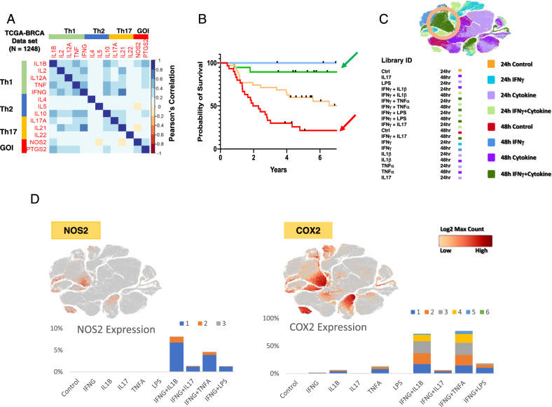
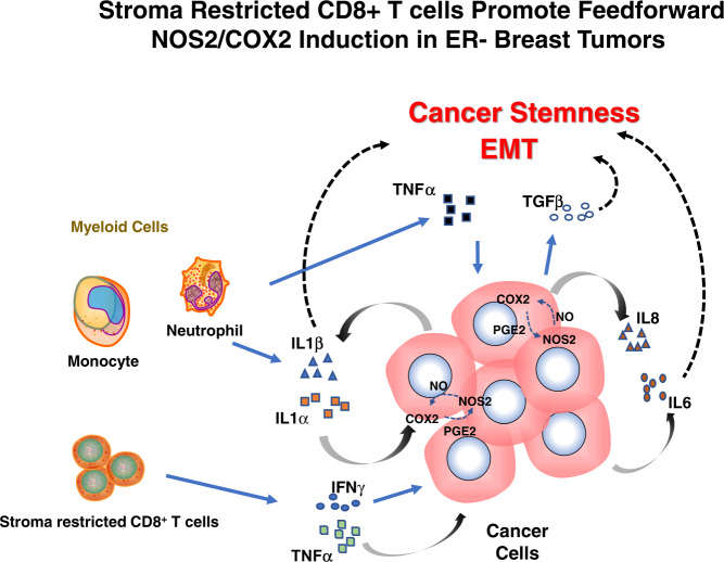
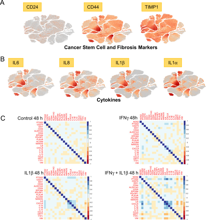
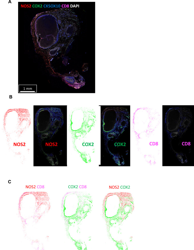
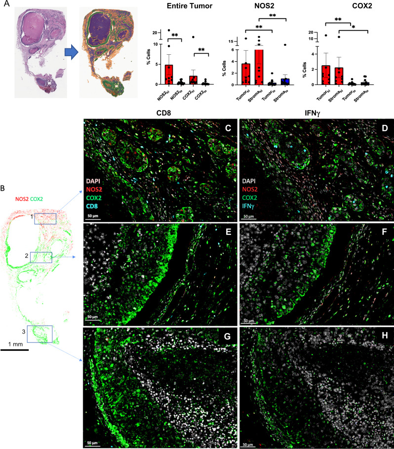
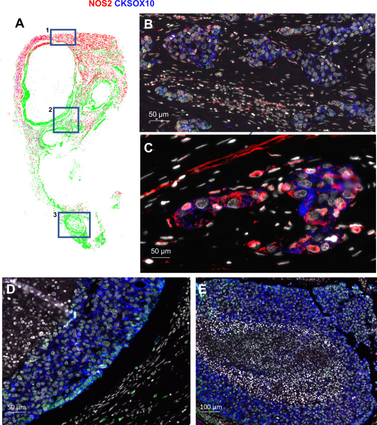
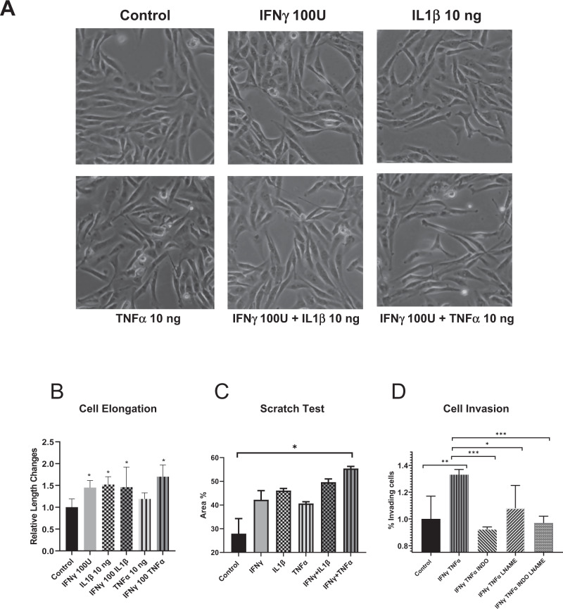
A strong correlation between NOS2 and COX2 tumor expression and poor clinical outcomes in ER breast cancer has been established. However, the mechanisms of tumor induction of these enzymes are unclear. Analysis of The Cancer Genome Atlas (TCGA) revealed correlations between NOS2 and COX2 expression and Th1 cytokines. Herein, single-cell RNAseq analysis of TNBC cells shows potent NOS2 and COX2 induction by IFNγ combined with IL1β or TNFα. Given that IFNγ is secreted by cytolytic lymphocytes, which improve clinical outcomes, this role of IFNγ presents a dichotomy. To explore this conundrum, tumor NOS2, COX2, and CD8 T cells were spatially analyzed in aggressive ER-, TNBC, and HER2 + breast tumors. High expression and clustering of NOS2-expressing tumor cells occurred at the tumor/stroma interface in the presence of stroma-restricted CD8 T cells. High expression and clustering of COX2-expressing tumor cells extended into immune desert regions in the tumor core where CD8 T cell penetration was limited or absent. Moreover, high NOS2-expressing tumor cells were proximal to areas with increased satellitosis, suggestive of cell clusters with a higher metastatic potential. Further in vitro experiments revealed that IFNγ + IL1β/TNFα increased the elongation and migration of treated tumor cells. This spatial analysis of the tumor microenvironment provides important insight into distinct neighborhoods where stroma-restricted CD8 T cells exist proximal to NOS2-expressing tumor niches that could have increased metastatic potential.
NOS2 和 COX2 肿瘤表达与 ER 阳性乳腺癌不良临床结局之间存在很强的相关性已得到确立。然而,这些酶诱导肿瘤的机制尚不清楚。对癌症基因组图谱(TCGA)的分析显示,NOS2 和 COX2 的表达与 Th1 细胞因子之间存在相关性。在此,对三阴性乳腺癌(TNBC)细胞的单细胞 RNAseq 分析表明,IFNγ 与 IL1β 或 TNFα 联合可强力诱导 NOS2 和 COX2 的表达。鉴于 IFNγ 是由改善临床结局的细胞毒性淋巴细胞分泌的,因此 IFNγ 的这种作用呈现出一种二分法。为了探索这一难题,在侵袭性 ER-、TNBC 和 HER2+乳腺癌肿瘤中对肿瘤 NOS2、COX2 和 CD8 T 细胞进行了空间分析。在存在限制于基质的 CD8 T 细胞的情况下,NOS2 表达的肿瘤细胞在肿瘤/基质界面高度表达和聚集。COX2 表达的肿瘤细胞在肿瘤核心的免疫荒漠区域高度表达和聚集,在这些区域,CD8 T 细胞的穿透受到限制或不存在。此外,高表达 NOS2 的肿瘤细胞靠近卫星现象增加的区域,提示细胞簇具有更高的转移潜力。进一步的体外实验表明,IFNγ+IL1β/TNFα 增加了经处理的肿瘤细胞的伸长和迁移。对肿瘤微环境的这种空间分析为存在于限制于基质的 CD8 T 细胞附近的 NOS2 表达肿瘤生态位提供了重要的见解,这些生态位可能具有更高的转移潜力。