Motolese Francesco, Lanzone Jacopo, Todisco Antonio, Rossi Mariagrazia, Santoro Francesca, Cruciani Alessandro, Capone Fioravante, Di Lazzaro Vincenzo, Pilato Fabio
Department of Medicine and Surgery, Unit of Neurology, Neurophysiology, Neurobiology and Psichiatry, Università Campus Bio-Medico di Roma, Rome, Italy.
Fondazione Policlinico Universitario Campus Bio-Medico, Rome, Italy.
Front Neurol. 2023 Apr 27;14:1178408. doi: 10.3389/fneur.2023.1178408. eCollection 2023.
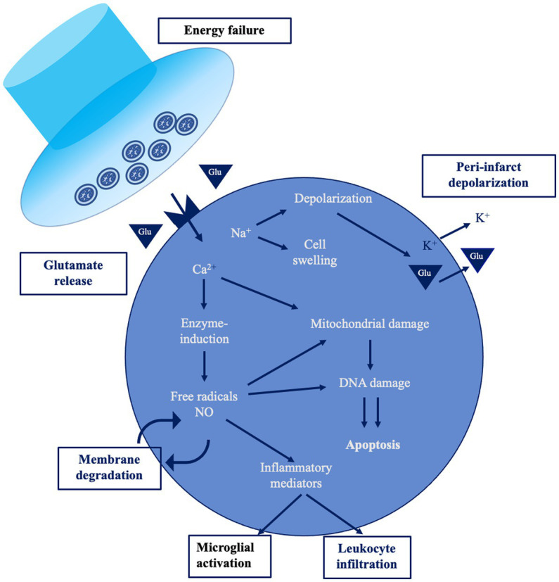
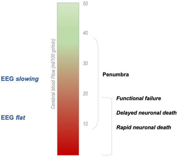
Ischemic stroke is characterized by a complex cascade of events starting from vessel occlusion. The term "penumbra" denotes the area of severely hypo-perfused brain tissue surrounding the ischemic core that can be potentially recovered if blood flow is reestablished. From the neurophysiological perspective, there are local alterations-reflecting the loss of function of the core and the penumbra-and widespread changes in neural networks functioning, since structural and functional connectivity is disrupted. These dynamic changes are closely related to blood flow in the affected area. However, the pathological process of stroke does not end after the acute phase, but it determines a long-term cascade of events, including changes of cortical excitability, that are quite precocious and might precede clinical evolution. Neurophysiological tools-such as Transcranial Magnetic Stimulation (TMS) or Electroencephalography (EEG)-have enough time resolution to efficiently reflect the pathological changes occurring after stroke. Even if they do not have a role in acute stroke management, EEG and TMS might be helpful for monitoring ischemia evolution-also in the sub-acute and chronic stages. The present review aims to describe the changes occurring in the infarcted area after stroke from the neurophysiological perspective, starting from the acute to the chronic phase.
缺血性中风的特征是从血管阻塞开始的一系列复杂事件。“半暗带”一词指的是围绕缺血核心的严重灌注不足的脑组织区域,如果血流得以恢复,该区域有可能恢复。从神经生理学角度来看,存在局部改变——反映核心区和半暗带功能丧失——以及神经网络功能的广泛变化,因为结构和功能连接被破坏。这些动态变化与受影响区域的血流密切相关。然而,中风的病理过程在急性期后并未结束,而是决定了一系列长期事件,包括皮质兴奋性的变化,这些变化相当早熟,可能先于临床进展。神经生理学工具,如经颅磁刺激(TMS)或脑电图(EEG),具有足够的时间分辨率,能够有效反映中风后发生的病理变化。即使它们在急性中风管理中不起作用,EEG和TMS可能有助于监测缺血进展,也适用于亚急性和慢性阶段。本综述旨在从神经生理学角度描述中风后梗死区域从急性期到慢性期发生的变化。