Walenski Matthew, Chen Yufen, Litcofsky Kaitlyn A, Caplan David, Kiran Swathi, Rapp Brenda, Parrish Todd B, Thompson Cynthia K
Department of Communication Sciences and Disorders, East Carolina University, Greenville, NC.
Center for the Neurobiology of Language Recovery, Northwestern University, Evanston, IL.
Neurobiol Lang (Camb). 2022 May 11;3(2):345-363. doi: 10.1162/nol_a_00068. eCollection 2022 May.
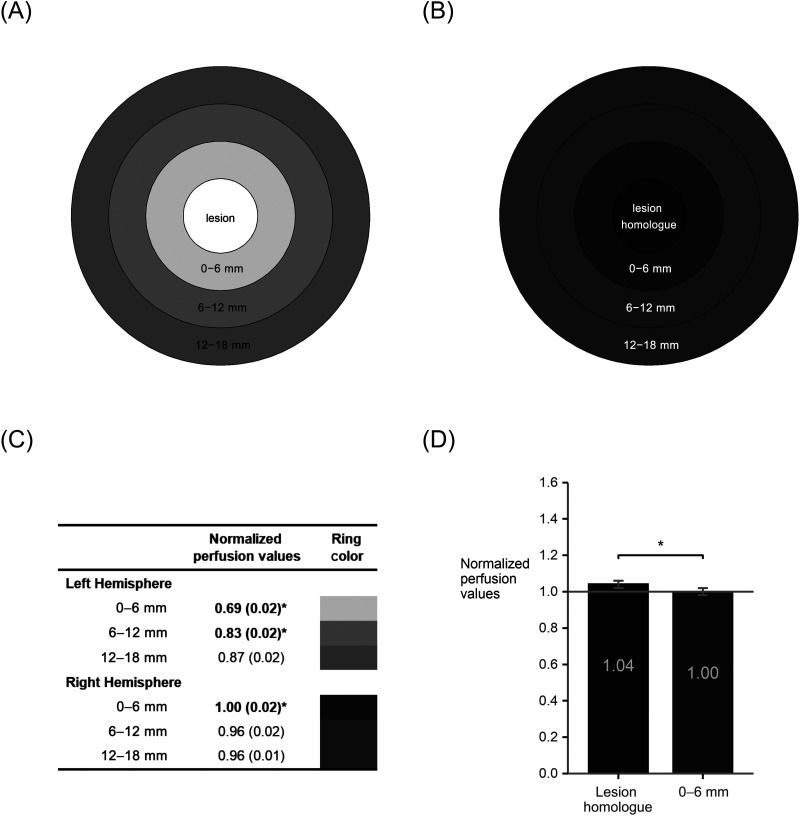
Stroke-induced alterations in cerebral blood flow (perfusion) may contribute to functional language impairments in chronic aphasia, particularly in perilesional tissue. Abnormal perfusion in this region may also serve as a biomarker for predicting functional improvements with behavioral treatment interventions. Using pseudo-continuous arterial spin labeling in magnetic resonance imaging (MRI), we examined perfusion in chronic aphasia, in perilesional rings in the left hemisphere and their right hemisphere homologues. In the left hemisphere we found a gradient pattern of decreasing perfusion closer to the lesion. The opposite pattern was found in the right hemisphere, with significantly increased perfusion close to the lesion homologue. Perfusion was also increased in the right hemisphere lesion homologue region relative to the surrounding tissue. We next examined changes in perfusion in two groups: one group who underwent MRI scanning before and after three months of a behavioral treatment intervention that led to significant language gains, and a second group who was scanned twice at a three-month interval without a treatment intervention. For both groups, there was no difference in perfusion over time in either the left or the right hemisphere. Moreover, within the treatment group pre-treatment perfusion scores did not predict treatment response; neither did pre-treatment perfusion predict post-treatment language performance. These results indicate that perfusion is chronically abnormal in both hemispheres, but chronically abnormal perfusion did not change in response to our behavioral treatment interventions, and did not predict responsiveness to language treatment.
中风引起的脑血流(灌注)改变可能导致慢性失语症患者出现功能性语言障碍,尤其是在病灶周围组织。该区域的异常灌注也可作为一种生物标志物,用于预测行为治疗干预后的功能改善情况。我们利用磁共振成像(MRI)中的伪连续动脉自旋标记技术,研究了慢性失语症患者左半球病灶周围环及其右半球对应区域的灌注情况。在左半球,我们发现靠近病灶处灌注呈递减的梯度模式。在右半球则发现相反的模式,靠近病灶对应区域的灌注显著增加。相对于周围组织,右半球病灶对应区域的灌注也有所增加。接下来,我们研究了两组患者的灌注变化:一组在接受为期三个月的行为治疗干预(该干预导致语言能力显著提高)前后进行了MRI扫描,另一组在三个月的间隔内进行了两次扫描,但未接受治疗干预。对于两组患者,左半球或右半球的灌注随时间均无差异。此外,在治疗组中,治疗前的灌注评分无法预测治疗反应;治疗前的灌注也无法预测治疗后的语言表现。这些结果表明,两个半球的灌注长期异常,但这种长期异常的灌注并未因我们的行为治疗干预而改变,也无法预测对语言治疗的反应。