• 文献检索
  • 文档翻译
  • 深度研究
  • 学术资讯
  • Suppr Zotero 插件Zotero 插件
  • 邀请有礼
  • 套餐&价格
  • 历史记录
应用&插件
Suppr Zotero 插件Zotero 插件浏览器插件Mac 客户端Windows 客户端微信小程序
定价
高级版会员购买积分包购买API积分包
服务
文献检索文档翻译深度研究API 文档MCP 服务
关于我们
关于 Suppr公司介绍联系我们用户协议隐私条款
关注我们

Suppr 超能文献

核心技术专利:CN118964589B侵权必究
粤ICP备2023148730 号-1Suppr @ 2026

文献检索

告别复杂PubMed语法,用中文像聊天一样搜索,搜遍4000万医学文献。AI智能推荐,让科研检索更轻松。

立即免费搜索

文件翻译

保留排版,准确专业,支持PDF/Word/PPT等文件格式,支持 12+语言互译。

免费翻译文档

深度研究

AI帮你快速写综述,25分钟生成高质量综述,智能提取关键信息,辅助科研写作。

立即免费体验

TCGA多形性胶质母细胞瘤队列中肿瘤异质性的生物标志物

Biomarkers of Tumor Heterogeneity in Glioblastoma Multiforme Cohort of TCGA.

作者信息

Winkelmaier Garrett, Koch Brandon, Bogardus Skylar, Borowsky Alexander D, Parvin Bahram

机构信息

Department of Electrical and Biomedical Engineering, College of Engineering, University of Nevada Reno, 1664 N. Virginia St., Reno, NV 89509, USA.

Department of Biostatics, College of Public Health, Ohio State University, 281 W. Lane Ave., Columbus, OH 43210, USA.

出版信息

Cancers (Basel). 2023 Apr 20;15(8):2387. doi: 10.3390/cancers15082387.

DOI:10.3390/cancers15082387
PMID:37190318
原文链接:https://pmc.ncbi.nlm.nih.gov/articles/PMC10137245/
Abstract

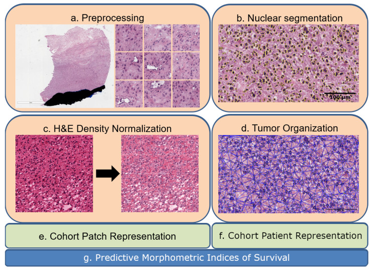
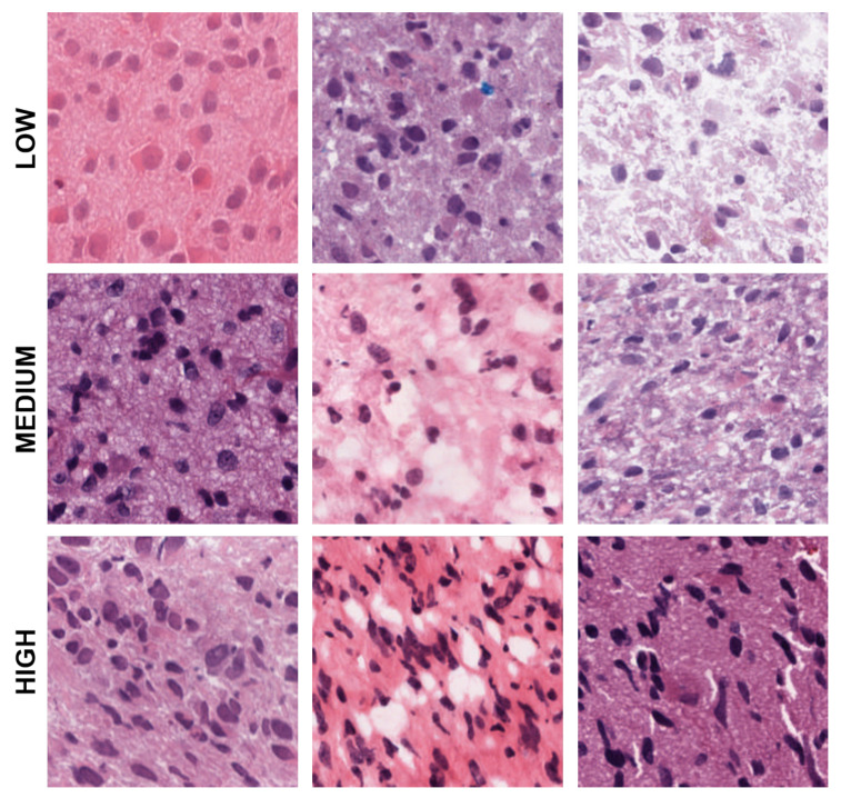
Tumor Whole Slide Images (WSI) are often heterogeneous, which hinders the discovery of biomarkers in the presence of confounding clinical factors. In this study, we present a pipeline for identifying biomarkers from the Glioblastoma Multiforme (GBM) cohort of WSIs from TCGA archive. The GBM cohort endures many technical artifacts while the discovery of GBM biomarkers is challenged because "age" is the single most confounding factor for predicting outcomes. The proposed approach relies on interpretable features (e.g., nuclear morphometric indices), effective similarity metrics for heterogeneity analysis, and robust statistics for identifying biomarkers. The pipeline first removes artifacts (e.g., pen marks) and partitions each WSI into patches for nuclear segmentation via an extended U-Net for subsequent quantitative representation. Given the variations in fixation and staining that can artificially modulate hematoxylin optical density (HOD), we extended Navab's Lab method to normalize images and reduce the impact of batch effects. The heterogeneity of each WSI is then represented either as probability density functions (PDF) per patient or as the composition of a dictionary predicted from the entire cohort of WSIs. For PDF- or dictionary-based methods, morphometric subtypes are constructed based on distances computed from optimal transport and linkage analysis or consensus clustering with Euclidean distances, respectively. For each inferred subtype, Kaplan-Meier and/or the Cox regression model are used to regress the survival time. Since age is the single most important confounder for predicting survival in GBM and there is an observed violation of the proportionality assumption in the Cox model, we use both age and age-squared coupled with the Likelihood ratio test and forest plots for evaluating competing statistics. Next, the PDF- and dictionary-based methods are combined to identify biomarkers that are predictive of survival. The combined model has the advantage of integrating global (e.g., cohort scale) and local (e.g., patient scale) attributes of morphometric heterogeneity, coupled with robust statistics, to reveal stable biomarkers. The results indicate that, after normalization of the GBM cohort, mean HOD, eccentricity, and cellularity are predictive of survival. Finally, we also stratified the GBM cohort as a function of EGFR expression and published genomic subtypes to reveal genomic-dependent morphometric biomarkers.

摘要

肿瘤全切片图像(WSI)通常具有异质性,这在存在混杂临床因素的情况下阻碍了生物标志物的发现。在本研究中,我们提出了一种从TCGA存档的胶质母细胞瘤(GBM)队列的WSI中识别生物标志物的流程。GBM队列存在许多技术伪像,而GBM生物标志物的发现面临挑战,因为“年龄”是预测结果的最主要混杂因素。所提出的方法依赖于可解释的特征(如核形态计量指标)、用于异质性分析的有效相似性度量以及用于识别生物标志物的稳健统计方法。该流程首先去除伪像(如笔触标记),并通过扩展的U-Net将每个WSI划分为用于核分割的图像块,以便进行后续的定量表征。鉴于固定和染色的差异会人为调节苏木精光密度(HOD),我们扩展了纳瓦布的实验室方法来对图像进行归一化,并减少批次效应的影响。然后,每个WSI的异质性要么表示为每个患者的概率密度函数(PDF),要么表示为从整个WSI队列预测的字典的组成。对于基于PDF或字典的方法,形态计量亚型分别基于从最优传输和连锁分析计算的距离或使用欧几里得距离的一致性聚类来构建。对于每个推断出的亚型,使用Kaplan-Meier和/或Cox回归模型对生存时间进行回归分析。由于年龄是预测GBM生存的最重要混杂因素,并且在Cox模型中观察到比例假设不成立,我们使用年龄和年龄平方,并结合似然比检验和森林图来评估竞争统计量。接下来,将基于PDF和字典的方法结合起来识别预测生存的生物标志物。组合模型具有整合形态计量异质性的全局(如队列规模)和局部(如患者规模)属性以及稳健统计量的优势,以揭示稳定的生物标志物。结果表明,在对GBM队列进行归一化后,平均HOD、偏心率和细胞密度可预测生存。最后,我们还根据表皮生长因子受体(EGFR)表达和已发表的基因组亚型对GBM队列进行分层,以揭示基因组依赖性形态计量生物标志物。

https://cdn.ncbi.nlm.nih.gov/pmc/blobs/3f09/10137245/909cef0e992a/cancers-15-02387-g010.jpg
https://cdn.ncbi.nlm.nih.gov/pmc/blobs/3f09/10137245/7a50b8a6f48e/cancers-15-02387-g001.jpg
https://cdn.ncbi.nlm.nih.gov/pmc/blobs/3f09/10137245/009c213c3aff/cancers-15-02387-g002.jpg
https://cdn.ncbi.nlm.nih.gov/pmc/blobs/3f09/10137245/aff8c279456b/cancers-15-02387-g003.jpg
https://cdn.ncbi.nlm.nih.gov/pmc/blobs/3f09/10137245/312b37553e16/cancers-15-02387-g004.jpg
https://cdn.ncbi.nlm.nih.gov/pmc/blobs/3f09/10137245/3efbee87f524/cancers-15-02387-g005.jpg
https://cdn.ncbi.nlm.nih.gov/pmc/blobs/3f09/10137245/eebfc79ccb8f/cancers-15-02387-g006.jpg
https://cdn.ncbi.nlm.nih.gov/pmc/blobs/3f09/10137245/87fc9e04b3b6/cancers-15-02387-g007.jpg
https://cdn.ncbi.nlm.nih.gov/pmc/blobs/3f09/10137245/f613e643419f/cancers-15-02387-g008.jpg
https://cdn.ncbi.nlm.nih.gov/pmc/blobs/3f09/10137245/e2c20f0362ea/cancers-15-02387-g009.jpg
https://cdn.ncbi.nlm.nih.gov/pmc/blobs/3f09/10137245/909cef0e992a/cancers-15-02387-g010.jpg

相似文献

1
Biomarkers of Tumor Heterogeneity in Glioblastoma Multiforme Cohort of TCGA.TCGA多形性胶质母细胞瘤队列中肿瘤异质性的生物标志物
Cancers (Basel). 2023 Apr 20;15(8):2387. doi: 10.3390/cancers15082387.
2
Invariant delineation of nuclear architecture in glioblastoma multiforme for clinical and molecular association.胶质母细胞瘤多形性中核架构的不变性描绘及其与临床和分子的关联。
IEEE Trans Med Imaging. 2013 Apr;32(4):670-82. doi: 10.1109/TMI.2012.2231420. Epub 2012 Dec 4.
3
New insights for precision treatment of glioblastoma from analysis of single-cell lncRNA expression.从单细胞 lncRNA 表达分析中获得胶质母细胞瘤精准治疗的新见解。
J Cancer Res Clin Oncol. 2021 Jul;147(7):1881-1895. doi: 10.1007/s00432-021-03584-9. Epub 2021 Mar 11.
4
PHENOTYPIC CHARACTERIZATION OF BREAST INVASIVE CARCINOMA VIA TRANSFERABLE TISSUE MORPHOMETRIC PATTERNS LEARNED FROM GLIOBLASTOMA MULTIFORME.通过从多形性胶质母细胞瘤中习得的可转移组织形态计量模式对乳腺浸润性癌进行表型特征分析。
Proc IEEE Int Symp Biomed Imaging. 2016 Apr;2016:1025-1028. doi: 10.1109/ISBI.2016.7493440.
5
Integrative Analysis of Cellular Morphometric Context Reveals Clinically Relevant Signatures in Lower Grade Glioma.细胞形态测量背景的综合分析揭示了低级别胶质瘤中与临床相关的特征。
Med Image Comput Comput Assist Interv. 2016 Oct;9900:72-80. doi: 10.1007/978-3-319-46720-7_9. Epub 2016 Oct 2.
6
CLASSIFICATION OF TUMOR HISTOPATHOLOGY VIA SPARSE FEATURE LEARNING.基于稀疏特征学习的肿瘤组织病理学分类
Proc IEEE Int Symp Biomed Imaging. 2013 Apr;2013. doi: 10.1109/ISBI.2013.6556499.
7
Folic acid supplementation and malaria susceptibility and severity among people taking antifolate antimalarial drugs in endemic areas.在流行地区,服用抗叶酸抗疟药物的人群中,叶酸补充剂与疟疾易感性和严重程度的关系。
Cochrane Database Syst Rev. 2022 Feb 1;2(2022):CD014217. doi: 10.1002/14651858.CD014217.
8
Morphometic analysis of TCGA glioblastoma multiforme.TCGA 胶质母细胞瘤的形态计量分析。
BMC Bioinformatics. 2011 Dec 20;12:484. doi: 10.1186/1471-2105-12-484.
9
A quantitative study of shape descriptors from glioblastoma multiforme phenotypes for predicting survival outcome.一项关于多形性胶质母细胞瘤表型形状描述符预测生存结果的定量研究。
Br J Radiol. 2016 Dec;89(1068):20160575. doi: 10.1259/bjr.20160575. Epub 2016 Oct 26.
10
Multilayer outperforms single-layer slide scanning in AI-based classification of whole slide images with low-burden acid-fast mycobacteria (AFB).多层扫描优于单层扫描,可基于人工智能对低负担抗酸分枝杆菌(AFB)全玻片图像进行分类。
Comput Methods Programs Biomed. 2023 Jun;234:107518. doi: 10.1016/j.cmpb.2023.107518. Epub 2023 Mar 28.

引用本文的文献

1
Applications of artificial intelligence in the analysis of histopathology images of gliomas: a review.人工智能在胶质瘤组织病理学图像分析中的应用:综述
Npj Imaging. 2024 Jul 1;2(1):16. doi: 10.1038/s44303-024-00020-8.

本文引用的文献

1
CBTRUS Statistical Report: Primary Brain and Other Central Nervous System Tumors Diagnosed in the United States in 2014-2018.美国 2014-2018 年诊断的原发性脑和其他中枢神经系统肿瘤 CBTRUS 统计报告。
Neuro Oncol. 2021 Oct 5;23(12 Suppl 2):iii1-iii105. doi: 10.1093/neuonc/noab200.
2
An enhanced loss function simplifies the deep learning model for characterizing the 3D organoid models.增强的损失函数简化了用于描述 3D 类器官模型的深度学习模型。
Bioinformatics. 2021 Sep 29;37(18):3084-3085. doi: 10.1093/bioinformatics/btab120.
3
PyHIST: A Histological Image Segmentation Tool.
PyHIST:一个组织学图像分割工具。
PLoS Comput Biol. 2020 Oct 19;16(10):e1008349. doi: 10.1371/journal.pcbi.1008349. eCollection 2020 Oct.
4
An automatic nuclei segmentation method based on deep convolutional neural networks for histopathology images.一种基于深度卷积神经网络的组织病理学图像细胞核自动分割方法。
BMC Biomed Eng. 2019 Oct 17;1:24. doi: 10.1186/s42490-019-0026-8. eCollection 2019.
5
Pathomic Fusion: An Integrated Framework for Fusing Histopathology and Genomic Features for Cancer Diagnosis and Prognosis.病理融合:融合组织病理学和基因组特征用于癌症诊断和预后的综合框架。
IEEE Trans Med Imaging. 2022 Apr;41(4):757-770. doi: 10.1109/TMI.2020.3021387. Epub 2022 Apr 1.
6
Deep Adversarial Training for Multi-Organ Nuclei Segmentation in Histopathology Images.深度学习在病理图像多器官细胞核分割中的应用
IEEE Trans Med Imaging. 2020 Nov;39(11):3257-3267. doi: 10.1109/TMI.2019.2927182. Epub 2020 Oct 28.
7
A Novel MKL Method for GBM Prognosis Prediction by Integrating Histopathological Image and Multi-Omics Data.一种基于 MKL 方法的新型 GBM 预后预测模型,整合了组织病理学图像和多组学数据。
IEEE J Biomed Health Inform. 2020 Jan;24(1):171-179. doi: 10.1109/JBHI.2019.2898471. Epub 2019 Feb 11.
8
Fast ScanNet: Fast and Dense Analysis of Multi-Gigapixel Whole-Slide Images for Cancer Metastasis Detection.Fast ScanNet:用于癌症转移检测的多吉字节全切片图像的快速和密集分析。
IEEE Trans Med Imaging. 2019 Aug;38(8):1948-1958. doi: 10.1109/TMI.2019.2891305. Epub 2019 Jan 7.
9
Fusion of encoder-decoder deep networks improves delineation of multiple nuclear phenotypes.编码器-解码器深度网络融合提高了多种核表型的勾画能力。
BMC Bioinformatics. 2018 Aug 7;19(1):294. doi: 10.1186/s12859-018-2285-0.
10
Predicting cancer outcomes from histology and genomics using convolutional networks.使用卷积网络从组织学和基因组学预测癌症结局。
Proc Natl Acad Sci U S A. 2018 Mar 27;115(13):E2970-E2979. doi: 10.1073/pnas.1717139115. Epub 2018 Mar 12.