Palmer Megan, Seddon James A, van der Zalm Marieke M, Hesseling Anneke C, Goussard Pierre, Schaaf H Simon, Morrison Julie, van Ginneken Bram, Melendez Jaime, Walters Elisabetta, Murphy Keelin
Faculty of Medicine and Health Sciences, Department of Paediatrics and Child Health, Demond Tutu TB Centre, Stellenbosch University, Cape Town, South Africa.
Department of Infectious Disease, Imperial College London, London, United Kingdom.
PLOS Glob Public Health. 2023 May 16;3(5):e0001799. doi: 10.1371/journal.pgph.0001799. eCollection 2023.
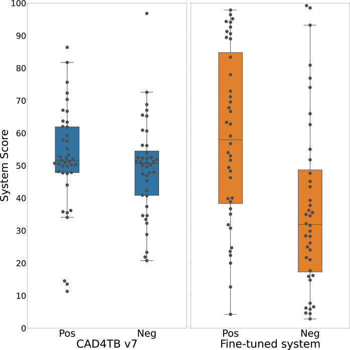
Diagnostic tools for paediatric tuberculosis remain limited, with heavy reliance on clinical algorithms which include chest x-ray. Computer aided detection (CAD) for tuberculosis on chest x-ray has shown promise in adults. We aimed to measure and optimise the performance of an adult CAD system, CAD4TB, to identify tuberculosis on chest x-rays from children with presumptive tuberculosis. Chest x-rays from 620 children <13 years enrolled in a prospective observational diagnostic study in South Africa, were evaluated. All chest x-rays were read by a panel of expert readers who attributed each with a radiological reference of either 'tuberculosis' or 'not tuberculosis'. Of the 525 chest x-rays included in this analysis, 80 (40 with a reference of 'tuberculosis' and 40 with 'not tuberculosis') were allocated to an independent test set. The remainder made up the training set. The performance of CAD4TB to identify 'tuberculosis' versus 'not tuberculosis' on chest x-ray against the radiological reference read was calculated. The CAD4TB software was then fine-tuned using the paediatric training set. We compared the performance of the fine-tuned model to the original model. Our findings were that the area under the receiver operating characteristic curve (AUC) of the original CAD4TB model, prior to fine-tuning, was 0.58. After fine-tuning there was an improvement in the AUC to 0.72 (p = 0.0016). In this first-ever description of the use of CAD to identify tuberculosis on chest x-ray in children, we demonstrate a significant improvement in the performance of CAD4TB after fine-tuning with a set of well-characterised paediatric chest x-rays. CAD has the potential to be a useful additional diagnostic tool for paediatric tuberculosis. We recommend replicating the methods we describe using a larger chest x-ray dataset from a more diverse population and evaluating the potential role of CAD to replace a human-read chest x-ray within treatment-decision algorithms for paediatric tuberculosis.
儿科结核病的诊断工具仍然有限,严重依赖包括胸部X光在内的临床算法。胸部X光计算机辅助检测(CAD)在成人结核病诊断中已显示出前景。我们旨在测量并优化成人CAD系统CAD4TB在识别疑似结核病儿童胸部X光片中结核病方面的性能。对南非一项前瞻性观察性诊断研究中纳入的620名13岁以下儿童的胸部X光片进行了评估。所有胸部X光片均由一组专家阅片者阅读,他们将每张片子归类为“结核病”或“非结核病”的放射学参考诊断。在本次分析纳入的525张胸部X光片中,80张(40张参考诊断为“结核病”,40张参考诊断为“非结核病”)被分配到一个独立测试集。其余片子组成训练集。计算了CAD4TB在胸部X光片上根据阅片者的放射学参考诊断来识别“结核病”与“非结核病”的性能。然后使用儿科训练集对CAD4TB软件进行微调。我们将微调后模型的性能与原始模型进行了比较。我们的研究结果是,在微调之前,原始CAD4TB模型的受试者工作特征曲线(AUC)下面积为0.58。微调后,AUC提高到了0.72(p = 0.0016)。在首次描述使用CAD识别儿童胸部X光片中结核病的研究中,我们证明了使用一组特征明确的儿科胸部X光片进行微调后,CAD4TB的性能有显著提高。CAD有可能成为儿科结核病一种有用的辅助诊断工具。我们建议使用来自更多样化人群的更大胸部X光数据集复制我们所描述的方法,并评估CAD在儿科结核病治疗决策算法中替代人工阅读胸部X光片的潜在作用。