Breuninger Marianne, van Ginneken Bram, Philipsen Rick H H M, Mhimbira Francis, Hella Jerry J, Lwilla Fred, van den Hombergh Jan, Ross Amanda, Jugheli Levan, Wagner Dirk, Reither Klaus
Swiss Tropical and Public Health Institute, Basel, Switzerland; Ifakara Health Institute, Bagamoyo, United Republic of Tanzania; Center for Infectious Diseases and Travel Medicine, University Hospital Freiburg, Freiburg, Germany.
Diagnostic Image Analysis Group, Radboud University Medical Center, Nijmegen, The Netherlands.
PLoS One. 2014 Sep 5;9(9):e106381. doi: 10.1371/journal.pone.0106381. eCollection 2014.
Chest radiography to diagnose and screen for pulmonary tuberculosis has limitations, especially due to inter-reader variability. Automating the interpretation has the potential to overcome this drawback and to deliver objective and reproducible results. The CAD4TB software is a computer-aided detection system that has shown promising preliminary findings. Evaluation studies in different settings are needed to assess diagnostic accuracy and practicability of use.
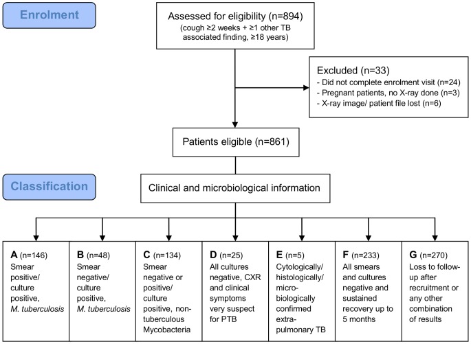
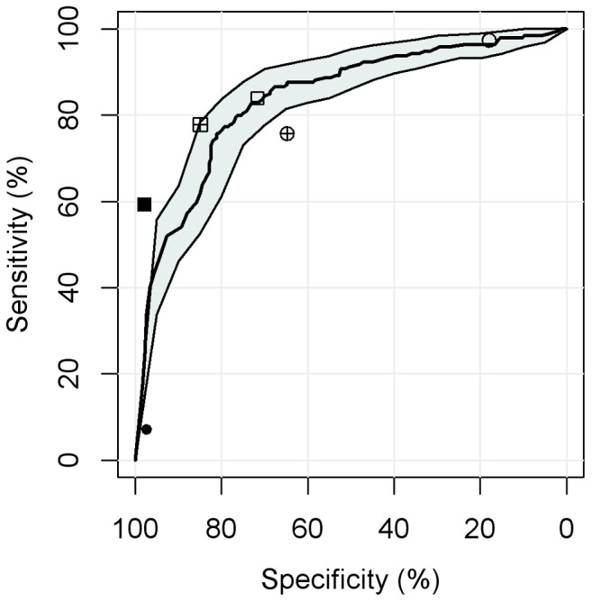
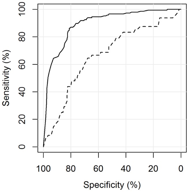
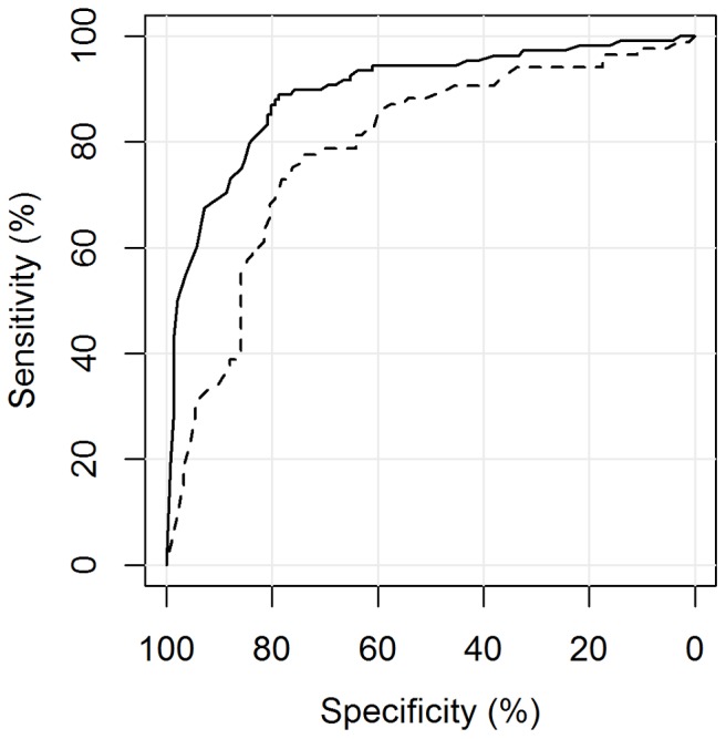
CAD4TB was evaluated on chest radiographs of patients with symptoms suggestive of pulmonary tuberculosis enrolled in two cohort studies in Tanzania. All patients were characterized by sputum smear microscopy and culture including subsequent antigen or molecular confirmation of Mycobacterium tuberculosis (M.tb) to determine the reference standard. Chest radiographs were read by the software and two human readers, one expert reader and one clinical officer. The sensitivity and specificity of CAD4TB was depicted using receiver operating characteristic (ROC) curves, the area under the curve calculated and the performance of the software compared to the results of human readers.
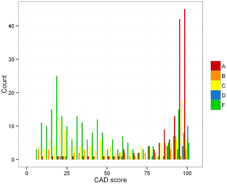
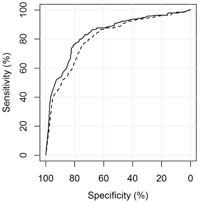
Of 861 study participants, 194 (23%) were culture-positive for M.tb. The area under the ROC curve of CAD4TB for the detection of culture-positive pulmonary tuberculosis was 0.84 (95% CI 0.80-0.88). CAD4TB was significantly more accurate for the discrimination of smear-positive cases against non TB patients than for smear-negative cases (p-value<0.01). It differentiated better between TB cases and non TB patients among HIV-negative compared to HIV-positive individuals (p<0.01). CAD4TB significantly outperformed the clinical officer, but did not reach the accuracy of the expert reader (p = 0.02), for a tuberculosis specific reading threshold.
CAD4TB accurately distinguished between the chest radiographs of culture-positive TB cases and controls. Further studies on cost-effectiveness, operational and ethical aspects should determine its place in diagnostic and screening algorithms.
胸部X线摄影用于诊断和筛查肺结核存在局限性,尤其是由于不同阅片者之间存在差异。实现解读自动化有可能克服这一缺点,并提供客观且可重复的结果。CAD4TB软件是一种计算机辅助检测系统,已显示出有前景的初步结果。需要在不同环境中进行评估研究,以评估其诊断准确性和实用性。
在坦桑尼亚的两项队列研究中,对有肺结核症状患者的胸部X线片进行CAD4TB评估。所有患者均通过痰涂片显微镜检查和培养进行特征分析,包括随后对结核分枝杆菌(M.tb)的抗原或分子确认,以确定参考标准。胸部X线片由该软件以及两名人类阅片者解读,一名专家阅片者和一名临床医生。使用受试者操作特征(ROC)曲线描绘CAD4TB的敏感性和特异性,计算曲线下面积,并将该软件的性能与人类阅片者的结果进行比较。
在861名研究参与者中,194人(23%)M.tb培养呈阳性。CAD4TB检测培养阳性肺结核的ROC曲线下面积为0.84(95%CI 0.80 - 0.88)。与涂片阴性病例相比,CAD4TB在区分涂片阳性病例与非结核患者方面的准确性显著更高(p值<0.01)。与HIV阳性个体相比,在HIV阴性个体中,CAD4TB在区分结核病例和非结核患者方面表现更好(p<0.01)。对于结核病特定阅读阈值,CAD4TB的表现显著优于临床医生,但未达到专家阅片者的准确性(p = 0.02)。
CAD4TB能够准确区分培养阳性结核病例和对照的胸部X线片。关于成本效益、操作和伦理方面的进一步研究应确定其在诊断和筛查算法中的地位。