Interdisciplinary Center Smell & Taste, Department of Otorhinolaryngology, Faculty of Medicine Carl Gustav Carus, Technische Universität Dresden, Fetscherstrasse 74, 01307, Dresden, Germany.
Eur Arch Otorhinolaryngol. 2023 Oct;280(10):4491-4499. doi: 10.1007/s00405-023-08019-4. Epub 2023 May 17.
In a previous neuroimaging study, patients with taste loss showed stronger activations in gustatory cortices compared to people with normal taste function during taste stimulations. The aim of the current study was to examine whether there are changes in central-nervous functional connectivity in patients with taste loss.
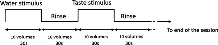
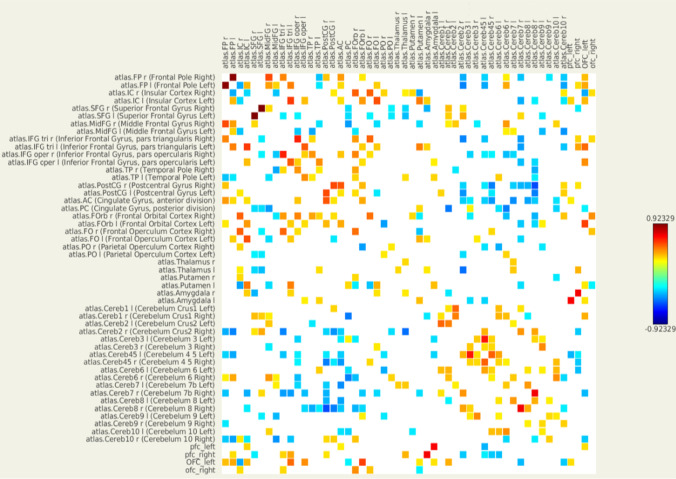
We selected 26 pairs of brain regions related to taste processing as our regions of interests (ROIs). Functional magnetic resonance imaging (fMRI) was used to measure brain responses in seven patients with taste loss and 12 healthy controls as they received taste stimulations (taste condition) and water (water condition). The data were analysed using ROI-to-ROI functional connectivity analysis (FCA).
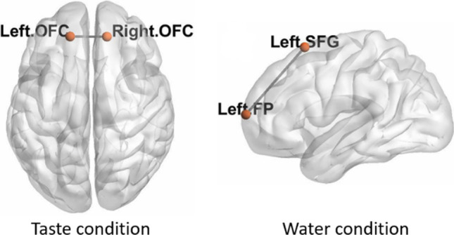
We observed weaker functional connectivity in the patient group between the left and right orbitofrontal cortex in the taste condition and between the left frontal pole and the left superior frontal gyrus in the water condition.
These results suggested that patients with taste loss experience changes of functional connectivity between brain regions not only relevant to taste processing but also to cognitive functions. While further studies are needed, fMRI might be helpful in diagnosing taste loss as an additional tool in exceptional cases.
在之前的一项神经影像学研究中,与味觉正常的人相比,味觉丧失的患者在味觉刺激期间,其味觉皮质的激活更强。本研究旨在探讨味觉丧失患者的中枢神经功能连接是否发生变化。
我们选择了 26 对与味觉处理相关的脑区作为感兴趣区(ROI)。使用功能磁共振成像(fMRI)测量了 7 名味觉丧失患者和 12 名健康对照者在接受味觉刺激(味觉条件)和水刺激(水条件)时的大脑反应。使用 ROI-ROI 功能连接分析(FCA)对数据进行分析。
我们发现,在味觉条件下,患者组的左侧和右侧眶额皮质之间以及在水条件下,左侧额极和左侧额上回之间的功能连接较弱。
这些结果表明,味觉丧失患者不仅在与味觉处理相关的脑区之间,而且在与认知功能相关的脑区之间经历了功能连接的变化。虽然还需要进一步的研究,但 fMRI 可能有助于作为特殊情况下的附加工具来诊断味觉丧失。