• 文献检索
  • 文档翻译
  • 深度研究
  • 学术资讯
  • Suppr Zotero 插件Zotero 插件
  • 邀请有礼
  • 套餐&价格
  • 历史记录
应用&插件
Suppr Zotero 插件Zotero 插件浏览器插件Mac 客户端Windows 客户端微信小程序
定价
高级版会员购买积分包购买API积分包
服务
文献检索文档翻译深度研究API 文档MCP 服务
关于我们
关于 Suppr公司介绍联系我们用户协议隐私条款
关注我们

Suppr 超能文献

核心技术专利:CN118964589B侵权必究
粤ICP备2023148730 号-1Suppr @ 2026

文献检索

告别复杂PubMed语法,用中文像聊天一样搜索,搜遍4000万医学文献。AI智能推荐,让科研检索更轻松。

立即免费搜索

文件翻译

保留排版,准确专业,支持PDF/Word/PPT等文件格式,支持 12+语言互译。

免费翻译文档

深度研究

AI帮你快速写综述,25分钟生成高质量综述,智能提取关键信息,辅助科研写作。

立即免费体验

使用时空映射校正多模态图像衍生的心脏几何结构中的偏差。

Correcting bias in cardiac geometries derived from multimodal images using spatiotemporal mapping.

机构信息

Auckland Bioengineering Institute, University of Auckland, 70 Symonds Street, Grafton, Auckland, 1010, New Zealand.

Department of Anatomy and Medical Imaging, University of Auckland, Auckland, New Zealand.

出版信息

Sci Rep. 2023 May 19;13(1):8118. doi: 10.1038/s41598-023-33968-5.

DOI:10.1038/s41598-023-33968-5
PMID:37208380
原文链接:https://pmc.ncbi.nlm.nih.gov/articles/PMC10199025/
Abstract

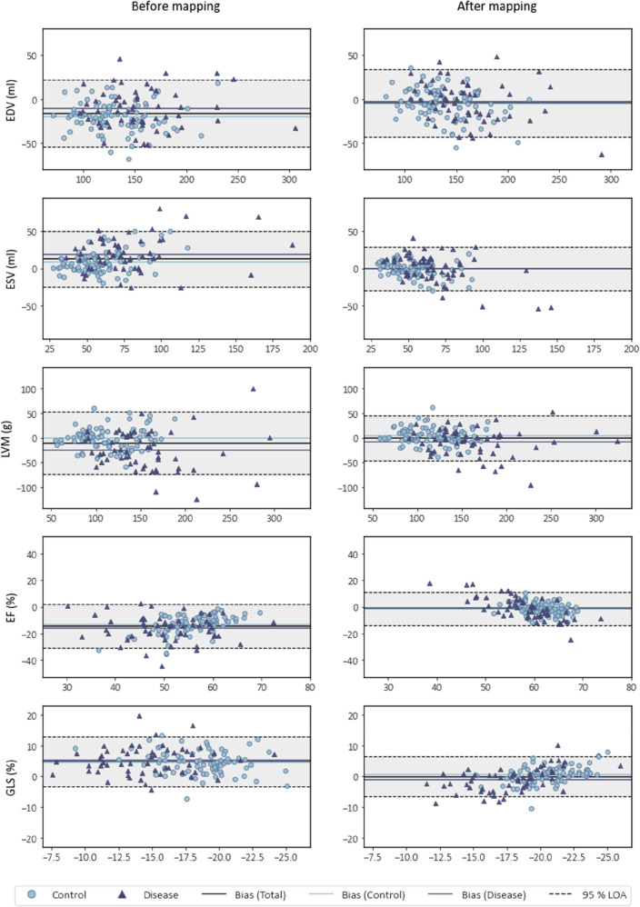
Cardiovascular imaging studies provide a multitude of structural and functional data to better understand disease mechanisms. While pooling data across studies enables more powerful and broader applications, performing quantitative comparisons across datasets with varying acquisition or analysis methods is problematic due to inherent measurement biases specific to each protocol. We show how dynamic time warping and partial least squares regression can be applied to effectively map between left ventricular geometries derived from different imaging modalities and analysis protocols to account for such differences. To demonstrate this method, paired real-time 3D echocardiography (3DE) and cardiac magnetic resonance (CMR) sequences from 138 subjects were used to construct a mapping function between the two modalities to correct for biases in left ventricular clinical cardiac indices, as well as regional shape. Leave-one-out cross-validation revealed a significant reduction in mean bias, narrower limits of agreement, and higher intraclass correlation coefficients for all functional indices between CMR and 3DE geometries after spatiotemporal mapping. Meanwhile, average root mean squared errors between surface coordinates of 3DE and CMR geometries across the cardiac cycle decreased from 7 ± 1 to 4 ± 1 mm for the total study population. Our generalised method for mapping between time-varying cardiac geometries obtained using different acquisition and analysis protocols enables the pooling of data between modalities and the potential for smaller studies to leverage large population databases for quantitative comparisons.

摘要

心血管成像研究提供了大量的结构和功能数据,以更好地了解疾病机制。虽然跨研究汇集数据可以实现更强大和更广泛的应用,但由于每个协议都存在特定的固有测量偏差,因此在具有不同采集或分析方法的数据集之间进行定量比较是有问题的。我们展示了如何应用动态时间扭曲和偏最小二乘回归来有效地在不同成像方式和分析协议得出的左心室几何形状之间进行映射,以解决这些差异。为了演示这种方法,使用 138 名受试者的实时 3 维超声心动图(3DE)和心脏磁共振(CMR)序列对配对数据进行了构建,以构建两种模式之间的映射函数,以校正左心室临床心脏指数和区域形状的偏差。在时空映射后,CMR 和 3DE 几何结构之间的所有功能指数的平均偏差、协议范围更窄,以及内部相关性系数都显著降低。同时,3DE 和 CMR 几何结构表面坐标在整个心动周期之间的平均均方根误差从 7±1 降至 4±1 毫米,适用于整个研究人群。我们用于不同采集和分析协议获得的时变心脏几何形状之间映射的通用方法,可以在模式之间汇集数据,并使较小的研究能够利用大型人群数据库进行定量比较。

https://cdn.ncbi.nlm.nih.gov/pmc/blobs/f6e8/10199025/837c9852b721/41598_2023_33968_Fig5_HTML.jpg
https://cdn.ncbi.nlm.nih.gov/pmc/blobs/f6e8/10199025/853fb56335db/41598_2023_33968_Fig1_HTML.jpg
https://cdn.ncbi.nlm.nih.gov/pmc/blobs/f6e8/10199025/c1ddef97f579/41598_2023_33968_Fig2_HTML.jpg
https://cdn.ncbi.nlm.nih.gov/pmc/blobs/f6e8/10199025/cc79f4299aa2/41598_2023_33968_Fig3_HTML.jpg
https://cdn.ncbi.nlm.nih.gov/pmc/blobs/f6e8/10199025/751878f54f6b/41598_2023_33968_Fig4_HTML.jpg
https://cdn.ncbi.nlm.nih.gov/pmc/blobs/f6e8/10199025/837c9852b721/41598_2023_33968_Fig5_HTML.jpg

相似文献

1
Correcting bias in cardiac geometries derived from multimodal images using spatiotemporal mapping.使用时空映射校正多模态图像衍生的心脏几何结构中的偏差。
Sci Rep. 2023 May 19;13(1):8118. doi: 10.1038/s41598-023-33968-5.
2
Feasibility of New Transthoracic Three-Dimensional Echocardiographic Automated Software for Left Heart Chamber Quantification in Children.新型经胸三维超声心动图自动软件用于儿童左心腔定量的可行性。
J Am Soc Echocardiogr. 2019 Jan;32(1):121-134.e1. doi: 10.1016/j.echo.2018.08.001. Epub 2018 Sep 18.
3
Three-dimensional echocardiography of the athlete's heart: a comparison with cardiac magnetic resonance imaging.运动员心脏的三维超声心动图:与心脏磁共振成像的比较。
Int J Cardiovasc Imaging. 2023 Feb;39(2):295-306. doi: 10.1007/s10554-022-02726-5. Epub 2022 Sep 23.
4
Optimal rotational interval for 3-dimensional echocardiography data acquisition for rapid and accurate measurement of left ventricular function.用于快速准确测量左心室功能的三维超声心动图数据采集的最佳旋转间隔。
J Am Soc Echocardiogr. 2000 Aug;13(8):715-22. doi: 10.1067/mje.2000.104899.
5
Accuracy and reproducibility of right ventricular quantification in patients with pressure and volume overload using single-beat three-dimensional echocardiography.使用单搏三维超声心动图对压力和容量负荷过重患者右心室定量的准确性和可重复性。
J Am Soc Echocardiogr. 2015 Mar;28(3):363-74. doi: 10.1016/j.echo.2014.10.012. Epub 2014 Dec 10.
6
Novel Approach to Three-Dimensional Echocardiographic Quantification of Right Ventricular Volumes and Function from Focused Views.从焦点视图评估右心室容积和功能的三维超声心动图新方法。
J Am Soc Echocardiogr. 2015 Oct;28(10):1222-31. doi: 10.1016/j.echo.2015.06.013. Epub 2015 Aug 1.
7
Feasibility and Accuracy of Automated Software for Transthoracic Three-Dimensional Left Ventricular Volume and Function Analysis: Comparisons with Two-Dimensional Echocardiography, Three-Dimensional Transthoracic Manual Method, and Cardiac Magnetic Resonance Imaging.自动化软件用于经胸三维左心室容量和功能分析的可行性和准确性:与二维超声心动图、三维经胸手动方法和心脏磁共振成像的比较。
J Am Soc Echocardiogr. 2017 Nov;30(11):1049-1058. doi: 10.1016/j.echo.2017.06.026. Epub 2017 Sep 12.
8
2D and 3D Echocardiography-Derived Indices of Left Ventricular Function and Shape: Relationship With Mortality.二维和三维超声心动图左心室功能和形态指标与死亡率的关系。
JACC Cardiovasc Imaging. 2018 Nov;11(11):1569-1579. doi: 10.1016/j.jcmg.2017.08.023. Epub 2017 Nov 15.
9
Right ventricular remodelling in pulmonary arterial hypertension with three-dimensional echocardiography: comparison with cardiac magnetic resonance imaging.三维超声心动图评估肺动脉高压患者右心室重构:与心脏磁共振成像的比较
Eur J Echocardiogr. 2010 Jan;11(1):64-73. doi: 10.1093/ejechocard/jep169. Epub 2009 Nov 24.
10
Left ventricular volume and ejection fraction measurements by fully automated 3D echocardiography left chamber quantification software versus CMR: A systematic review and meta-analysis.全自动化 3D 超声心动图左心室定量软件与 CMR 测量左心室容积和射血分数的比较:系统评价和荟萃分析。
J Cardiol. 2023 Jan;81(1):19-25. doi: 10.1016/j.jjcc.2022.08.007. Epub 2022 Sep 2.

引用本文的文献

1
Assessment of knee flexion in young children with prosthetic knee components using dynamic time warping.使用动态时间规整技术评估佩戴假肢膝关节组件的幼儿的膝关节屈曲情况。
Front Rehabil Sci. 2023 Aug 25;4:1227870. doi: 10.3389/fresc.2023.1227870. eCollection 2023.

本文引用的文献

1
Reverse re-modelling chronic heart failure by reinstating heart rate variability.通过恢复心率变异性来逆转重塑慢性心力衰竭。
Basic Res Cardiol. 2022 Feb 1;117(1):4. doi: 10.1007/s00395-022-00911-0.
2
Influence of heart rate correction formulas on QTc interval stability.心率校正公式对 QTc 间期稳定性的影响。
Sci Rep. 2021 Jul 12;11(1):14269. doi: 10.1038/s41598-021-93774-9.
3
The SPARC DRC: Building a Resource for the Autonomic Nervous System Community.SPARC数据资源中心:为自主神经系统领域建立一个资源库。
Front Physiol. 2021 Jun 24;12:693735. doi: 10.3389/fphys.2021.693735. eCollection 2021.
4
Morphologies and prognostic significance of left ventricular volume/time curves with cardiac magnetic resonance in patients with non-ischaemic heart failure and left bundle branch block.左心室容积/时间曲线形态及其在非缺血性心力衰竭伴左束支传导阻滞患者心脏磁共振中的预后意义。
Int J Cardiovasc Imaging. 2021 Jul;37(7):2245-2255. doi: 10.1007/s10554-021-02194-3. Epub 2021 Feb 26.
5
Diagnosis of Left Ventricular Diastolic Dysfunction Using Cardiac Magnetic Resonance Imaging: Comparison of Volume-Time Curves Derived from Long- and Short-Axis Cine Steady-State Free Precession Datasets.应用心脏磁共振成像诊断左心室舒张功能障碍:长轴和短轴电影稳态自由进动数据集容积-时间曲线的比较。
Rofo. 2020 Aug;192(8):764-775. doi: 10.1055/a-1108-1892. Epub 2020 Feb 27.
6
Non-invasive prediction of genotype positive-phenotype negative in hypertrophic cardiomyopathy by 3D modern shape analysis.3D 现代形态分析对肥厚型心肌病基因型阳性表型阴性的无创预测。
Exp Physiol. 2019 Nov;104(11):1688-1700. doi: 10.1113/EP087551. Epub 2019 Sep 1.
7
Modeling left ventricular dynamics with characteristic deformation modes.用特征变形模式对左心室动力学进行建模。
Biomech Model Mechanobiol. 2019 Dec;18(6):1683-1696. doi: 10.1007/s10237-019-01168-8. Epub 2019 May 25.
8
Improving performance of 3D speckle tracking in arterial hypertension and paroxysmal atrial fibrillation by using novel strain parameters.利用新型应变参数提高动脉高血压和阵发性心房颤动的 3D 斑点追踪性能。
Sci Rep. 2019 May 14;9(1):7382. doi: 10.1038/s41598-019-43855-7.
9
Independent Left Ventricular Morphometric Atlases Show Consistent Relationships with Cardiovascular Risk Factors: A UK Biobank Study.独立的左心室形态计量学图谱与心血管危险因素具有一致的关系:一项英国生物库研究。
Sci Rep. 2019 Feb 4;9(1):1130. doi: 10.1038/s41598-018-37916-6.
10
Computational models in cardiology.心脏病学中的计算模型。
Nat Rev Cardiol. 2019 Feb;16(2):100-111. doi: 10.1038/s41569-018-0104-y.