TH-PPM Group, Physics Department, Faculty of Sciences, Beni-Suef University, Beni Suef, 62514, Egypt.
Applied College, Khamis Mushait, King Khalid University, Abha, 62529, Kingdom of Saudi Arabia.
Sci Rep. 2023 May 19;13(1):8115. doi: 10.1038/s41598-023-34601-1.
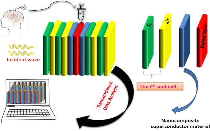
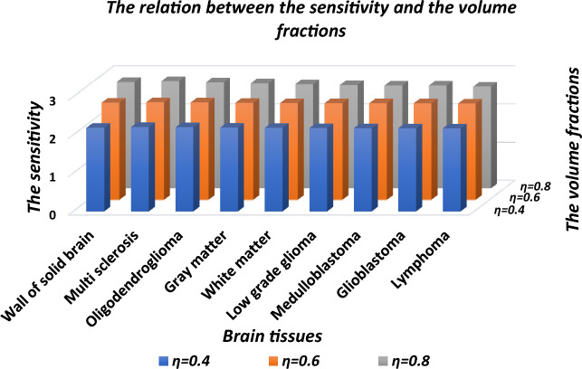
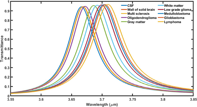
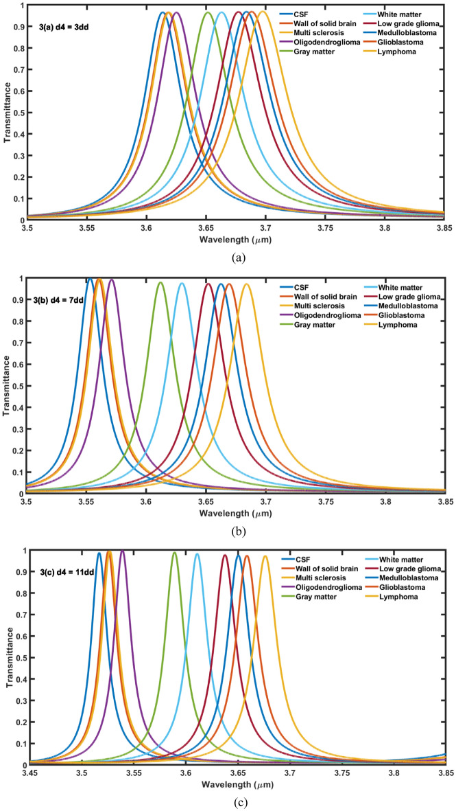
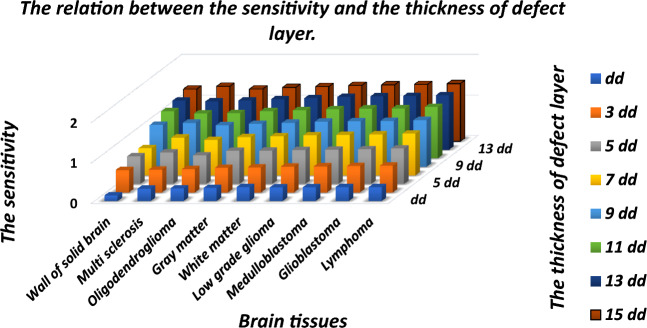
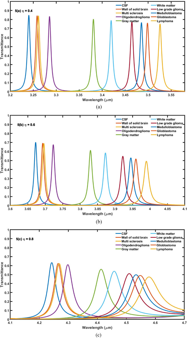
In the present research work we have theoretically examined the biosensing capabilities of proposed one dimensional defective photonic crystal for swift detection of malignant brain tissues. The transfer matrix formulation and MATLAB computational tool have been used to examine the transmission properties of proposed structure. The identical buffer layers of nanocomposite superconducting material have been used either side of cavity region to enhance the interaction between incident light and different brain tissue samples poured into the cavity region. All the investigations have been carried out under normal incidence to suppress the experimental liabilities involved. We have investigated the biosensing performance of the proposed design by changing the values of two internal parameters (1) the cavity layer thickness (d) and (2) volume fraction (η) of nanocomposite buffer layers one by one to get the optimum biosensing performance from the structure. It has been found that the sensitivity of the proposed design becomes 1.42607 μm/RIU when the cavity region of thickness 15dd is loaded with lymphoma brain tissue. This value of sensitivity can be further increased to 2.66136 μm/RIU with η = 0.8. The findings of this work are very beneficial for designing of various bio-sensing structures composed of nanocomposite materials of diversified biomedical applications.
在本研究工作中,我们从理论上研究了所提出的一维缺陷光子晶体的生物传感能力,以便快速检测恶性脑组织。使用传输矩阵公式和 MATLAB 计算工具来研究所提出结构的传输特性。在腔区域的两侧使用相同的纳米复合超导材料缓冲层,以增强进入腔区域的不同脑组织样本与入射光之间的相互作用。所有的研究都是在正常入射下进行的,以抑制涉及的实验责任。我们通过改变两个内部参数(1)腔层厚度(d)和(2)纳米复合缓冲层的体积分数(η)的值,对所提出的设计的生物传感性能进行了研究,以从结构中获得最佳的生物传感性能。当厚度为 15dd 的腔区域中填充淋巴瘤脑组织时,发现该设计的灵敏度变为 1.42607μm/RIU。通过将 η = 0.8,该灵敏度值可以进一步增加到 2.66136μm/RIU。这项工作的发现对于设计由各种生物医学应用的纳米复合材料组成的各种生物传感结构非常有益。