• 文献检索
  • 文档翻译
  • 深度研究
  • 学术资讯
  • Suppr Zotero 插件Zotero 插件
  • 邀请有礼
  • 套餐&价格
  • 历史记录
应用&插件
Suppr Zotero 插件Zotero 插件浏览器插件Mac 客户端Windows 客户端微信小程序
定价
高级版会员购买积分包购买API积分包
服务
文献检索文档翻译深度研究API 文档MCP 服务
关于我们
关于 Suppr公司介绍联系我们用户协议隐私条款
关注我们

Suppr 超能文献

核心技术专利:CN118964589B侵权必究
粤ICP备2023148730 号-1Suppr @ 2026

文献检索

告别复杂PubMed语法,用中文像聊天一样搜索,搜遍4000万医学文献。AI智能推荐,让科研检索更轻松。

立即免费搜索

文件翻译

保留排版,准确专业,支持PDF/Word/PPT等文件格式,支持 12+语言互译。

免费翻译文档

深度研究

AI帮你快速写综述,25分钟生成高质量综述,智能提取关键信息,辅助科研写作。

立即免费体验

Seg2Link:一种高效、通用的三维图像堆栈半自动细胞分割解决方案。

Seg2Link: an efficient and versatile solution for semi-automatic cell segmentation in 3D image stacks.

机构信息

Graduate School of Science, Nagoya City University, Nagoya, Japan.

RIKEN Center for Biosystems Dynamics Research, Kobe, Japan.

出版信息

Sci Rep. 2023 May 22;13(1):7109. doi: 10.1038/s41598-023-34232-6.

DOI:10.1038/s41598-023-34232-6
PMID:37217545
原文链接:https://pmc.ncbi.nlm.nih.gov/articles/PMC10203120/
Abstract

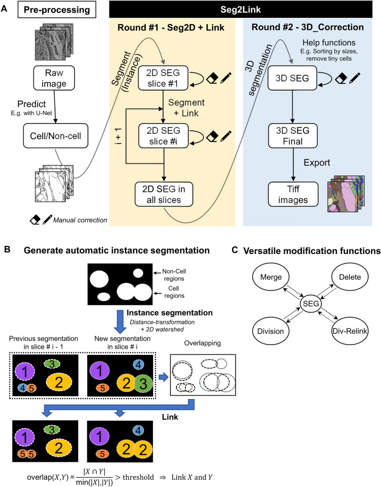
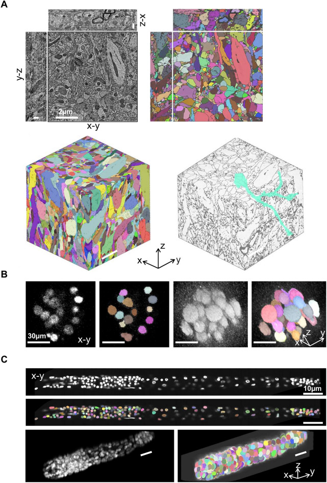
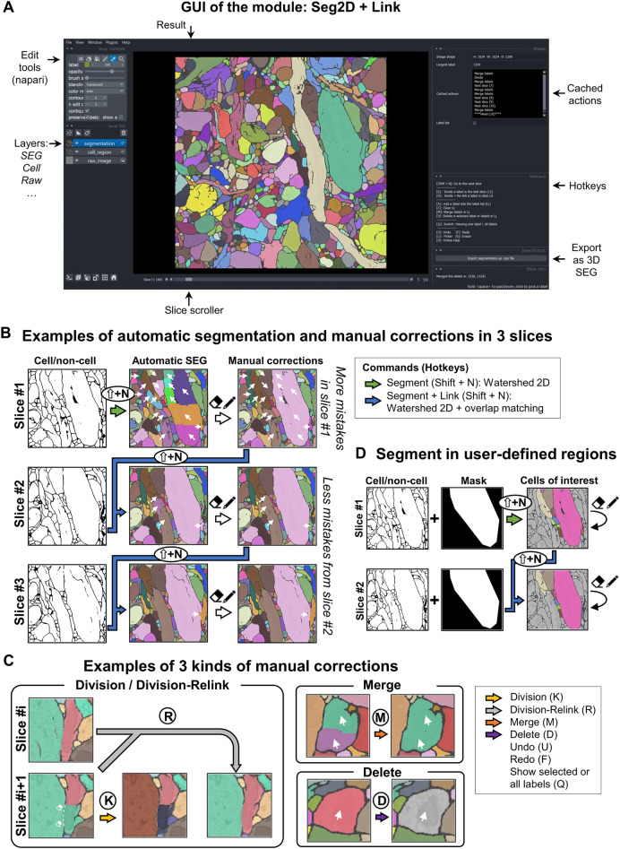
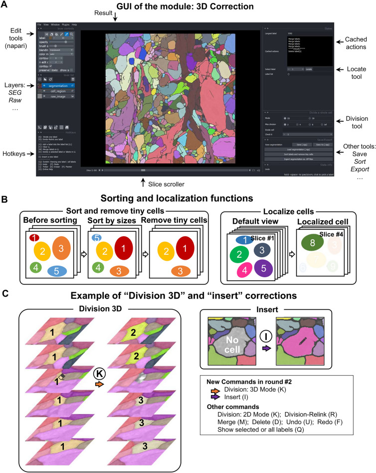
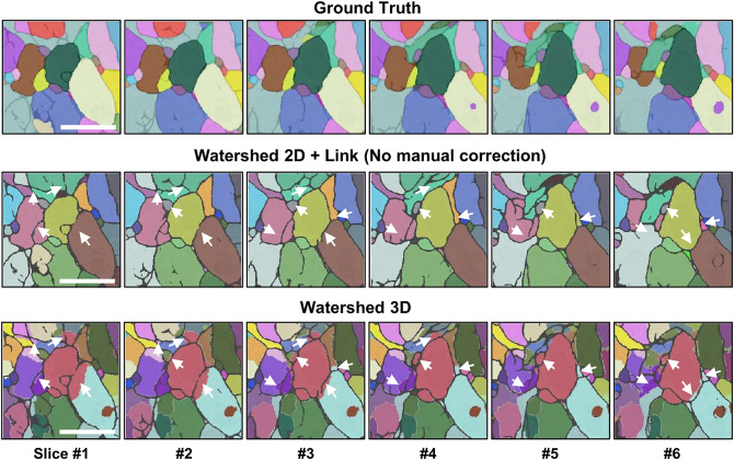
Recent advances in microscopy techniques, especially in electron microscopy, are transforming biomedical studies by acquiring large quantities of high-precision 3D cell image stacks. To examine cell morphology and connectivity in organs such as the brain, scientists need to conduct cell segmentation, which extracts individual cell regions of different shapes and sizes from a 3D image. This is challenging due to the indistinct images often encountered in real biomedical research: in many cases, automatic segmentation methods inevitably contain numerous mistakes in the segmentation results, even when using advanced deep learning methods. To analyze 3D cell images effectively, a semi-automated software solution is needed that combines powerful deep learning techniques with the ability to perform post-processing, generate accurate segmentations, and incorporate manual corrections. To address this gap, we developed Seg2Link, which takes deep learning predictions as inputs and use watershed 2D + cross-slice linking to generate more accurate automatic segmentations than previous methods. Additionally, it provides various manual correction tools essential for correcting mistakes in 3D segmentation results. Moreover, our software has been optimized for efficiently processing large 3D images in diverse organisms. Thus, Seg2Link offers an practical solution for scientists to study cell morphology and connectivity in 3D image stacks.

摘要

近年来,显微镜技术,特别是电子显微镜技术的进步,通过获取大量高精度的 3D 细胞图像堆栈,正在改变生物医学研究。为了研究大脑等器官中的细胞形态和连接性,科学家需要进行细胞分割,即从 3D 图像中提取出不同形状和大小的单个细胞区域。这是一项具有挑战性的工作,因为在实际的生物医学研究中经常会遇到不清晰的图像:在许多情况下,即使使用先进的深度学习方法,自动分割方法也不可避免地会在分割结果中包含许多错误。为了有效地分析 3D 细胞图像,需要一种结合强大的深度学习技术和后处理能力的半自动软件解决方案,以生成更准确的自动分割,并结合手动校正。为了解决这一差距,我们开发了 Seg2Link,它将深度学习预测作为输入,并使用分水岭 2D+跨切片链接生成比以前的方法更准确的自动分割。此外,它还提供了各种手动校正工具,对于校正 3D 分割结果中的错误至关重要。此外,我们的软件经过优化,可以有效地处理来自不同生物体的大型 3D 图像。因此,Seg2Link 为科学家研究 3D 图像堆栈中的细胞形态和连接性提供了一种实用的解决方案。

https://cdn.ncbi.nlm.nih.gov/pmc/blobs/c3a9/10203120/848c994933cd/41598_2023_34232_Fig5_HTML.jpg
https://cdn.ncbi.nlm.nih.gov/pmc/blobs/c3a9/10203120/e0016fad68ff/41598_2023_34232_Fig1_HTML.jpg
https://cdn.ncbi.nlm.nih.gov/pmc/blobs/c3a9/10203120/f5751a873c75/41598_2023_34232_Fig2_HTML.jpg
https://cdn.ncbi.nlm.nih.gov/pmc/blobs/c3a9/10203120/5a688281b6dc/41598_2023_34232_Fig3_HTML.jpg
https://cdn.ncbi.nlm.nih.gov/pmc/blobs/c3a9/10203120/b2a701e2e363/41598_2023_34232_Fig4_HTML.jpg
https://cdn.ncbi.nlm.nih.gov/pmc/blobs/c3a9/10203120/848c994933cd/41598_2023_34232_Fig5_HTML.jpg

相似文献

1
Seg2Link: an efficient and versatile solution for semi-automatic cell segmentation in 3D image stacks.Seg2Link:一种高效、通用的三维图像堆栈半自动细胞分割解决方案。
Sci Rep. 2023 May 22;13(1):7109. doi: 10.1038/s41598-023-34232-6.
2
Semi-automatic stitching of filamentous structures in image stacks from serial-section electron tomography.序列切片电子断层扫描图像堆栈中丝状结构的半自动拼接。
J Microsc. 2021 Oct;284(1):25-44. doi: 10.1111/jmi.13039. Epub 2021 Jul 9.
3
Bi-channel image registration and deep-learning segmentation (BIRDS) for efficient, versatile 3D mapping of mouse brain.双通道图像配准和深度学习分割(BIRDS)用于高效、通用的小鼠大脑 3D 映射。
Elife. 2021 Jan 18;10:e63455. doi: 10.7554/eLife.63455.
4
VAST (Volume Annotation and Segmentation Tool): Efficient Manual and Semi-Automatic Labeling of Large 3D Image Stacks.VAST(体积注释和分割工具):高效的大型 3D 图像堆栈手动和半自动标注。
Front Neural Circuits. 2018 Oct 16;12:88. doi: 10.3389/fncir.2018.00088. eCollection 2018.
5
A method for the evaluation of thousands of automated 3D stem cell segmentations.一种评估数千个自动化3D干细胞分割结果的方法。
J Microsc. 2015 Dec;260(3):363-76. doi: 10.1111/jmi.12303. Epub 2015 Aug 13.
6
An analytical tool that quantifies cellular morphology changes from three-dimensional fluorescence images.一种可对三维荧光图像中的细胞形态变化进行量化的分析工具。
J Vis Exp. 2012 Aug 31(66):e4233. doi: 10.3791/4233.
7
Single-cell segmentation in bacterial biofilms with an optimized deep learning method enables tracking of cell lineages and measurements of growth rates.利用优化的深度学习方法对细菌生物膜中的单细胞进行分割,可实现细胞谱系追踪和生长速率测量。
Mol Microbiol. 2023 Jun;119(6):659-676. doi: 10.1111/mmi.15064. Epub 2023 Apr 17.
8
3D dento-maxillary osteolytic lesion and active contour segmentation pilot study in CBCT: semi-automatic vs manual methods.CBCT中3D牙颌面溶骨性病变与活动轮廓分割的初步研究:半自动与手动方法对比
Dentomaxillofac Radiol. 2015;44(8):20150079. doi: 10.1259/dmfr.20150079. Epub 2015 May 21.
9
Rapid and Semi-automated Extraction of Neuronal Cell Bodies and Nuclei from Electron Microscopy Image Stacks.从电子显微镜图像堆栈中快速半自动提取神经元细胞体和细胞核
Methods Mol Biol. 2016;1427:277-90. doi: 10.1007/978-1-4939-3615-1_16.
10
Automated subcortical volume estimation from 2D MRI in epilepsy and implications for clinical trials.从癫痫的 2D MRI 中自动进行皮质下体积估算及其对临床试验的影响。
Neuroradiology. 2022 May;64(5):935-947. doi: 10.1007/s00234-021-02811-x. Epub 2021 Oct 18.

引用本文的文献

1
Sparse Annotation is Sufficient for Bootstrapping Dense Segmentation.稀疏标注足以用于引导密集分割。
Res Sq. 2024 Nov 14:rs.3.rs-5339143. doi: 10.21203/rs.3.rs-5339143/v1.
2
Sparse Annotation is Sufficient for Bootstrapping Dense Segmentation.稀疏标注足以用于引导密集分割。
bioRxiv. 2024 Oct 26:2024.06.14.599135. doi: 10.1101/2024.06.14.599135.
3
Neuraminidase inhibition promotes the collective migration of neurons and recovery of brain function.神经氨酸酶抑制促进神经元的集体迁移和大脑功能的恢复。

本文引用的文献

1
TrackMate 7: integrating state-of-the-art segmentation algorithms into tracking pipelines.TrackMate 7:将最先进的分割算法集成到跟踪管道中。
Nat Methods. 2022 Jul;19(7):829-832. doi: 10.1038/s41592-022-01507-1. Epub 2022 Jun 2.
2
Regulation of liver subcellular architecture controls metabolic homeostasis.调控肝亚细胞结构控制代谢稳态。
Nature. 2022 Mar;603(7902):736-742. doi: 10.1038/s41586-022-04488-5. Epub 2022 Mar 9.
3
Image Segmentation Using Deep Learning: A Survey.基于深度学习的图像分割技术综述。
EMBO Mol Med. 2024 Jun;16(6):1228-1253. doi: 10.1038/s44321-024-00073-7. Epub 2024 May 24.
IEEE Trans Pattern Anal Mach Intell. 2022 Jul;44(7):3523-3542. doi: 10.1109/TPAMI.2021.3059968. Epub 2022 Jun 3.
4
Cellpose: a generalist algorithm for cellular segmentation.Cellpose:一种通用的细胞分割算法。
Nat Methods. 2021 Jan;18(1):100-106. doi: 10.1038/s41592-020-01018-x. Epub 2020 Dec 14.
5
3D convolutional neural networks-based segmentation to acquire quantitative criteria of the nucleus during mouse embryogenesis.基于 3D 卷积神经网络的分割技术,获取小鼠胚胎发生过程中细胞核的定量标准。
NPJ Syst Biol Appl. 2020 Oct 20;6(1):32. doi: 10.1038/s41540-020-00152-8.
6
A connectome and analysis of the adult central brain.一个成年中枢大脑的连接组和分析。
Elife. 2020 Sep 7;9:e57443. doi: 10.7554/eLife.57443.
7
Is It Time to Start Transitioning From 2D to 3D Cell Culture?是时候开始从二维细胞培养向三维细胞培养转变了吗?
Front Mol Biosci. 2020 Mar 6;7:33. doi: 10.3389/fmolb.2020.00033. eCollection 2020.
8
UNI-EM: An Environment for Deep Neural Network-Based Automated Segmentation of Neuronal Electron Microscopic Images.UNI-EM:一种基于深度神经网络的神经元电子显微镜图像自动分割环境。
Sci Rep. 2019 Dec 19;9(1):19413. doi: 10.1038/s41598-019-55431-0.
9
Dynamic Changes in Ultrastructure of the Primary Cilium in Migrating Neuroblasts in the Postnatal Brain.出生后大脑中迁移神经母细胞的初级纤毛超微结构的动态变化。
J Neurosci. 2019 Dec 11;39(50):9967-9988. doi: 10.1523/JNEUROSCI.1503-19.2019. Epub 2019 Nov 4.
10
ilastik: interactive machine learning for (bio)image analysis.ilastik:用于(生物)图像处理的交互式机器学习。
Nat Methods. 2019 Dec;16(12):1226-1232. doi: 10.1038/s41592-019-0582-9. Epub 2019 Sep 30.