• 文献检索
  • 文档翻译
  • 深度研究
  • 学术资讯
  • Suppr Zotero 插件Zotero 插件
  • 邀请有礼
  • 套餐&价格
  • 历史记录
应用&插件
Suppr Zotero 插件Zotero 插件浏览器插件Mac 客户端Windows 客户端微信小程序
定价
高级版会员购买积分包购买API积分包
服务
文献检索文档翻译深度研究API 文档MCP 服务
关于我们
关于 Suppr公司介绍联系我们用户协议隐私条款
关注我们

Suppr 超能文献

核心技术专利:CN118964589B侵权必究
粤ICP备2023148730 号-1Suppr @ 2026

文献检索

告别复杂PubMed语法,用中文像聊天一样搜索,搜遍4000万医学文献。AI智能推荐,让科研检索更轻松。

立即免费搜索

文件翻译

保留排版,准确专业,支持PDF/Word/PPT等文件格式,支持 12+语言互译。

免费翻译文档

深度研究

AI帮你快速写综述,25分钟生成高质量综述,智能提取关键信息,辅助科研写作。

立即免费体验

ACCT 是一个快速且易于使用的自动细胞计数工具,它使用机器学习进行 2D 图像分割。

ACCT is a fast and accessible automatic cell counting tool using machine learning for 2D image segmentation.

机构信息

Graduate Program of Genetics, Genomics and Bioinformatics, University of California, Riverside, Riverside, CA, 92521, USA.

School of Medicine, Division of Biomedical Sciences, University of California, Riverside, Riverside, CA, 92521, USA.

出版信息

Sci Rep. 2023 May 22;13(1):8213. doi: 10.1038/s41598-023-34943-w.

DOI:10.1038/s41598-023-34943-w
PMID:37217558
原文链接:https://pmc.ncbi.nlm.nih.gov/articles/PMC10202925/
Abstract

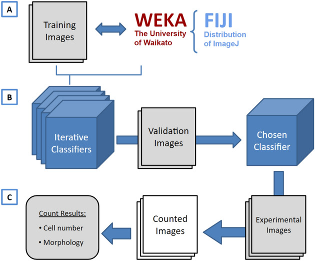
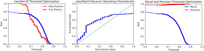
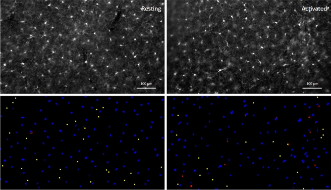
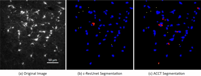
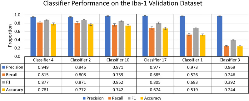
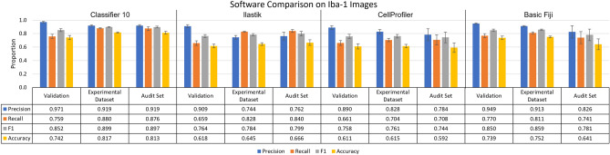
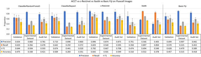
Counting cells is a cornerstone of tracking disease progression in neuroscience. A common approach for this process is having trained researchers individually select and count cells within an image, which is not only difficult to standardize but also very time-consuming. While tools exist to automatically count cells in images, the accuracy and accessibility of such tools can be improved. Thus, we introduce a novel tool ACCT: Automatic Cell Counting with Trainable Weka Segmentation which allows for flexible automatic cell counting via object segmentation after user-driven training. ACCT is demonstrated with a comparative analysis of publicly available images of neurons and an in-house dataset of immunofluorescence-stained microglia cells. For comparison, both datasets were manually counted to demonstrate the applicability of ACCT as an accessible means to automatically quantify cells in a precise manner without the need for computing clusters or advanced data preparation.

摘要

细胞计数是神经科学中跟踪疾病进展的基础。一种常见的方法是让经过训练的研究人员单独选择并计数图像中的细胞,这种方法不仅难以标准化,而且非常耗时。虽然有工具可以自动计数图像中的细胞,但这些工具的准确性和可访问性可以进一步提高。因此,我们引入了一种新的工具 ACCT:使用可训练的 WEKA 分割进行自动细胞计数,该工具允许在用户驱动的培训后通过对象分割进行灵活的自动细胞计数。ACCT 通过对神经元的公开图像和免疫荧光染色的小胶质细胞的内部数据集进行比较分析来进行演示。为了进行比较,两个数据集都进行了手动计数,以证明 ACCT 是一种可访问的方法,可以自动以精确的方式定量细胞,而无需计算聚类或进行高级数据准备。

https://cdn.ncbi.nlm.nih.gov/pmc/blobs/651e/10202925/a6a96f0a65d5/41598_2023_34943_Fig9_HTML.jpg
https://cdn.ncbi.nlm.nih.gov/pmc/blobs/651e/10202925/50f5ec8933f4/41598_2023_34943_Fig1_HTML.jpg
https://cdn.ncbi.nlm.nih.gov/pmc/blobs/651e/10202925/d5feeaae027c/41598_2023_34943_Fig2_HTML.jpg
https://cdn.ncbi.nlm.nih.gov/pmc/blobs/651e/10202925/1f8cf01aae36/41598_2023_34943_Fig3_HTML.jpg
https://cdn.ncbi.nlm.nih.gov/pmc/blobs/651e/10202925/88438b8a669a/41598_2023_34943_Fig4_HTML.jpg
https://cdn.ncbi.nlm.nih.gov/pmc/blobs/651e/10202925/41dfdf8f83eb/41598_2023_34943_Fig5_HTML.jpg
https://cdn.ncbi.nlm.nih.gov/pmc/blobs/651e/10202925/22983de6e609/41598_2023_34943_Fig6_HTML.jpg
https://cdn.ncbi.nlm.nih.gov/pmc/blobs/651e/10202925/cf2dbf6be1eb/41598_2023_34943_Fig7_HTML.jpg
https://cdn.ncbi.nlm.nih.gov/pmc/blobs/651e/10202925/a545452b0da0/41598_2023_34943_Fig8_HTML.jpg
https://cdn.ncbi.nlm.nih.gov/pmc/blobs/651e/10202925/a6a96f0a65d5/41598_2023_34943_Fig9_HTML.jpg

相似文献

1
ACCT is a fast and accessible automatic cell counting tool using machine learning for 2D image segmentation.ACCT 是一个快速且易于使用的自动细胞计数工具,它使用机器学习进行 2D 图像分割。
Sci Rep. 2023 May 22;13(1):8213. doi: 10.1038/s41598-023-34943-w.
2
Comparison of two automatic cell-counting solutions for fluorescent microscopic images.两种用于荧光显微镜图像的自动细胞计数解决方案的比较。
J Microsc. 2015 Oct;260(1):107-16. doi: 10.1111/jmi.12272. Epub 2015 Jun 22.
3
Trainable Weka Segmentation: a machine learning tool for microscopy pixel classification.可训练的 WEKA 分割:一种用于显微镜像素分类的机器学习工具。
Bioinformatics. 2017 Aug 1;33(15):2424-2426. doi: 10.1093/bioinformatics/btx180.
4
Automatic ground truth for deep learning stereology of immunostained neurons and microglia in mouse neocortex.深度学习免疫染色神经元和小胶质细胞体视学的自动地面实况。
J Chem Neuroanat. 2019 Jul;98:1-7. doi: 10.1016/j.jchemneu.2019.02.006. Epub 2019 Mar 2.
5
Nuclear-labeling index analysis (NLIA), a software package used to perform accurate automation of cell nuclear-labeling index analysis on immunohistochemically stained rat liver samples.核标记指数分析(NLIA),是一个用于对免疫组化染色的大鼠肝脏样本进行细胞核标记指数分析精确自动化操作的软件包。
Comput Methods Programs Biomed. 2000 Aug;63(1):55-70. doi: 10.1016/s0169-2607(00)00075-4.
6
Performance Comparison of Five Methods for Number Counting on the ImageJ Platform: Assessing the Built-in Tool and Machine-Learning-Based Extension.基于 ImageJ 平台的五种数字计数方法的性能比较:评估内置工具和基于机器学习的扩展。
Int J Mol Sci. 2022 May 26;23(11):6009. doi: 10.3390/ijms23116009.
7
A Cell Segmentation/Tracking Tool Based on Machine Learning.一种基于机器学习的细胞分割/追踪工具。
Methods Mol Biol. 2019;2040:399-422. doi: 10.1007/978-1-4939-9686-5_19.
8
Classifying changes in LN-18 glial cell morphology: a supervised machine learning approach to analyzing cell microscopy data via FIJI and WEKA.对 LN-18 神经胶质细胞形态变化进行分类:一种通过 FIJI 和 WEKA 对细胞显微镜数据进行分析的有监督机器学习方法。
Med Biol Eng Comput. 2020 Jul;58(7):1419-1430. doi: 10.1007/s11517-020-02177-x. Epub 2020 Apr 21.
9
An automatic image analysis approach to quantify stained cell cultures.一种用于量化染色细胞培养物的自动图像分析方法。
Cell Mol Biol (Noisy-le-grand). 2007 Apr 27;53(2):44-50.
10
Bi-channel image registration and deep-learning segmentation (BIRDS) for efficient, versatile 3D mapping of mouse brain.双通道图像配准和深度学习分割(BIRDS)用于高效、通用的小鼠大脑 3D 映射。
Elife. 2021 Jan 18;10:e63455. doi: 10.7554/eLife.63455.

引用本文的文献

1
β-Amyloid induces microglial expression of GPC4 and APOE leading to increased neuronal tau pathology and toxicity.β-淀粉样蛋白诱导小胶质细胞表达磷脂酰肌醇蛋白聚糖4(GPC4)和载脂蛋白E(APOE),导致神经元tau蛋白病变和毒性增加。
Mol Neurodegener. 2025 Aug 29;20(1):96. doi: 10.1186/s13024-025-00883-4.
2
Behavioural and neural correlates of social hierarchy formation in a sex-changing fish.一种性逆转鱼类社会等级形成的行为和神经关联
Proc Biol Sci. 2025 May;292(2046):20242097. doi: 10.1098/rspb.2024.2097. Epub 2025 May 14.
3
SEMTWIST Quantification of Biofilm Infection in Human Chronic Wound Using Scanning Electron Microscopy and Machine Learning.

本文引用的文献

1
Automating cell counting in fluorescent microscopy through deep learning with c-ResUnet.通过使用c-ResUnet深度学习实现荧光显微镜下细胞计数的自动化。
Sci Rep. 2021 Nov 25;11(1):22920. doi: 10.1038/s41598-021-01929-5.
2
CellProfiler 4: improvements in speed, utility and usability.CellProfiler 4:在速度、实用性和易用性方面的改进。
BMC Bioinformatics. 2021 Sep 10;22(1):433. doi: 10.1186/s12859-021-04344-9.
3
Array programming with NumPy.使用 NumPy 进行数组编程。
使用扫描电子显微镜和机器学习对人类慢性伤口生物膜感染进行SEMTWIST量化
Adv Wound Care (New Rochelle). 2025 Aug;14(8):393-408. doi: 10.1089/wound.2024.0291. Epub 2025 May 13.
4
Challenges of Cell Counting in Cell Therapy Products.细胞治疗产品细胞计数的挑战。
Cell Transplant. 2024 Jan-Dec;33:9636897241293628. doi: 10.1177/09636897241293628.
5
A semi-automated cell tracking protocol for quantitative analyses of neutrophil swarming to sterile and S. aureus contaminated bone implants in a mouse femur model.一种半自动的细胞追踪方案,用于定量分析中性粒细胞在无菌和金黄色葡萄球菌污染的小鼠股骨骨植入物模型中的迁移。
PLoS One. 2024 Jun 20;19(6):e0296140. doi: 10.1371/journal.pone.0296140. eCollection 2024.
6
PCP auto count: a novel Fiji/ImageJ plug-in for automated quantification of planar cell polarity and cell counting.PCP自动计数:一种用于平面细胞极性自动定量和细胞计数的新型斐济/ImageJ插件。
Front Cell Dev Biol. 2024 May 17;12:1394031. doi: 10.3389/fcell.2024.1394031. eCollection 2024.
7
PCP Auto Count: A Novel Fiji/ImageJ plug-in for automated quantification of planar cell polarity and cell counting.PCP自动计数:一种用于平面细胞极性自动定量和细胞计数的新型斐济/ImageJ插件。
bioRxiv. 2024 Feb 15:2024.01.30.578047. doi: 10.1101/2024.01.30.578047.
8
A semi-automated cell tracking protocol for quantitative analyses of neutrophil swarming to sterile and contaminated bone implants in a mouse femur model.一种用于在小鼠股骨模型中对中性粒细胞向无菌和污染骨植入物趋集进行定量分析的半自动细胞追踪方案。
bioRxiv. 2023 Dec 8:2023.12.07.570663. doi: 10.1101/2023.12.07.570663.
Nature. 2020 Sep;585(7825):357-362. doi: 10.1038/s41586-020-2649-2. Epub 2020 Sep 16.
4
A pivotal role for Interferon-α receptor-1 in neuronal injury induced by HIV-1.干扰素-α受体-1在 HIV-1 诱导的神经元损伤中的关键作用。
J Neuroinflammation. 2020 Jul 29;17(1):226. doi: 10.1186/s12974-020-01894-2.
5
SciPy 1.0: fundamental algorithms for scientific computing in Python.SciPy 1.0:Python 中的科学计算基础算法。
Nat Methods. 2020 Mar;17(3):261-272. doi: 10.1038/s41592-019-0686-2. Epub 2020 Feb 3.
6
ilastik: interactive machine learning for (bio)image analysis.ilastik:用于(生物)图像处理的交互式机器学习。
Nat Methods. 2019 Dec;16(12):1226-1232. doi: 10.1038/s41592-019-0582-9. Epub 2019 Sep 30.
7
Trainable Weka Segmentation: a machine learning tool for microscopy pixel classification.可训练的 WEKA 分割:一种用于显微镜像素分类的机器学习工具。
Bioinformatics. 2017 Aug 1;33(15):2424-2426. doi: 10.1093/bioinformatics/btx180.
8
CellProfiler Analyst: interactive data exploration, analysis and classification of large biological image sets.细胞图像分析软件(CellProfiler Analyst):用于大型生物图像集的交互式数据探索、分析与分类。
Bioinformatics. 2016 Oct 15;32(20):3210-3212. doi: 10.1093/bioinformatics/btw390. Epub 2016 Jun 26.
9
The search for true numbers of neurons and glial cells in the human brain: A review of 150 years of cell counting.探寻人类大脑中神经元和神经胶质细胞的真实数量:150年细胞计数研究综述
J Comp Neurol. 2016 Dec 15;524(18):3865-3895. doi: 10.1002/cne.24040. Epub 2016 Jun 16.
10
Microglial dynamics and role in the healthy and diseased brain: a paradigm of functional plasticity.小胶质细胞在健康和患病大脑中的动态变化及作用:功能可塑性范例
Neuroscientist. 2015 Apr;21(2):169-84. doi: 10.1177/1073858414530512. Epub 2014 Apr 10.