Division of Chronic Inflammation and Cancer, German Cancer Research Center Heidelberg (DKFZ), Heidelberg, Germany.
Division of Chronic Inflammation and Cancer, German Cancer Research Center Heidelberg (DKFZ), Heidelberg, Germany; Research Unit of Radiation Cytogenetics (ZYTO), Helmholtz Zentrum München, Neuherberg, Germany; Institute of Molecular Oncology and Functional Genomics, Clinic and Polyclinic for Internal Medicine II, Klinikum rechts der Isar of the Technical University of Munich (TUM), Munich, Germany; Translational Pancreatic Cancer Research Center, Clinic and Polyclinic for Internal Medicine II, Klinikum rechts der Isar of the Technical University of Munich (TUM), Munich, Germany.
J Hepatol. 2023 Aug;79(2):296-313. doi: 10.1016/j.jhep.2023.04.037. Epub 2023 May 22.
BACKGROUND & AIMS: The progression of non-alcoholic steatohepatitis (NASH) to fibrosis and hepatocellular carcinoma (HCC) is aggravated by auto-aggressive T cells. The gut-liver axis contributes to NASH, but the mechanisms involved and the consequences for NASH-induced fibrosis and liver cancer remain unknown. We investigated the role of gastrointestinal B cells in the development of NASH, fibrosis and NASH-induced HCC.
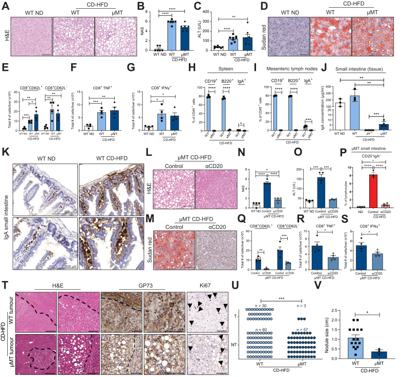
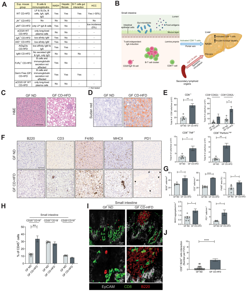
C57BL/6J wild-type (WT), B cell-deficient and different immunoglobulin-deficient or transgenic mice were fed distinct NASH-inducing diets or standard chow for 6 or 12 months, whereafter NASH, fibrosis, and NASH-induced HCC were assessed and analysed. Specific pathogen-free/germ-free WT and μMT mice (containing B cells only in the gastrointestinal tract) were fed a choline-deficient high-fat diet, and treated with an anti-CD20 antibody, whereafter NASH and fibrosis were assessed. Tissue biopsy samples from patients with simple steatosis, NASH and cirrhosis were analysed to correlate the secretion of immunoglobulins to clinicopathological features. Flow cytometry, immunohistochemistry and single-cell RNA-sequencing analysis were performed in liver and gastrointestinal tissue to characterise immune cells in mice and humans.
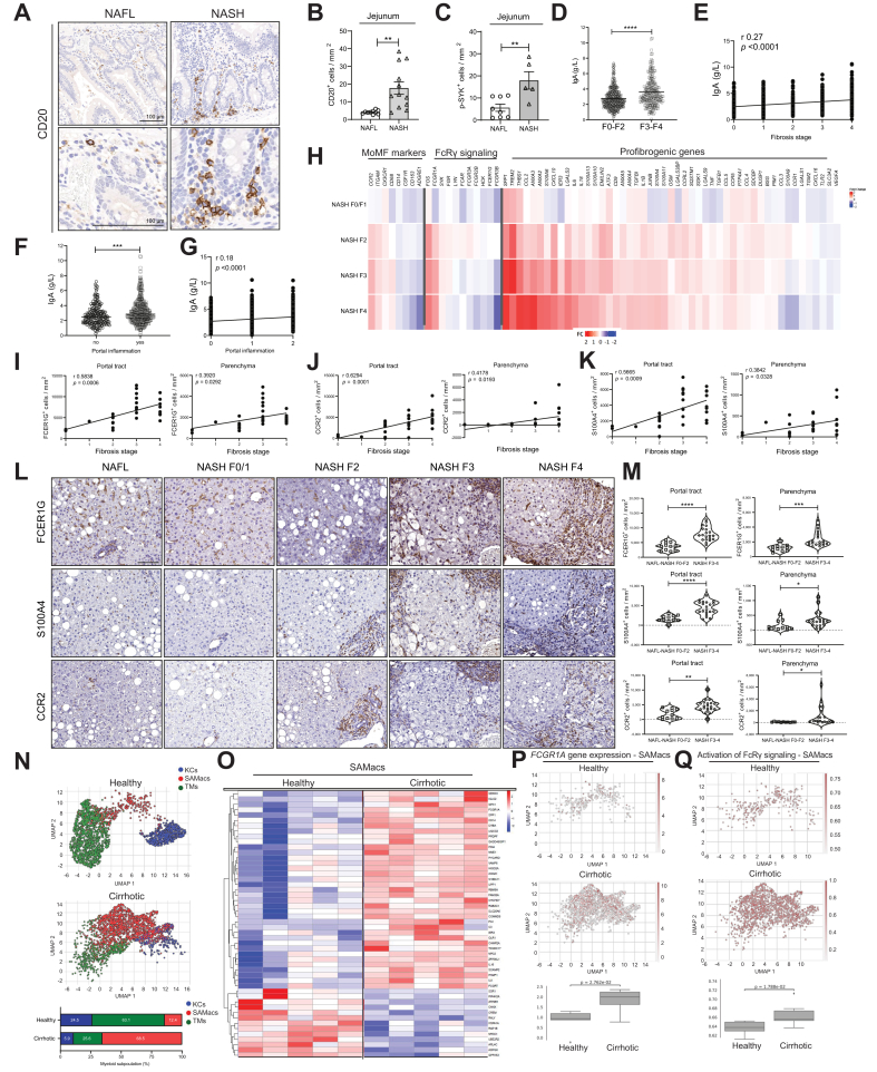
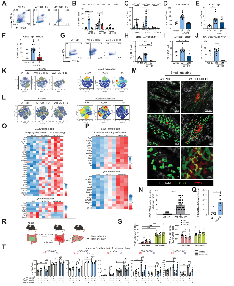
Activated intestinal B cells were increased in mouse and human NASH samples and licensed metabolic T-cell activation to induce NASH independently of antigen specificity and gut microbiota. Genetic or therapeutic depletion of systemic or gastrointestinal B cells prevented or reverted NASH and liver fibrosis. IgA secretion was necessary for fibrosis induction by activating CD11b+CCR2+F4/80+CD11c-FCGR1+ hepatic myeloid cells through an IgA-FcR signalling axis. Similarly, patients with NASH had increased numbers of activated intestinal B cells; additionally, we observed a positive correlation between IgA levels and activated FcRg+ hepatic myeloid cells, as well the extent of liver fibrosis.
Intestinal B cells and the IgA-FcR signalling axis represent potential therapeutic targets for the treatment of NASH.
There is currently no effective treatment for non-alcoholic steatohepatitis (NASH), which is associated with a substantial healthcare burden and is a growing risk factor for hepatocellular carcinoma (HCC). We have previously shown that NASH is an auto-aggressive condition aggravated, amongst others, by T cells. Therefore, we hypothesized that B cells might have a role in disease induction and progression. Our present work highlights that B cells have a dual role in NASH pathogenesis, being implicated in the activation of auto-aggressive T cells and the development of fibrosis via activation of monocyte-derived macrophages by secreted immunoglobulins (e.g., IgA). Furthermore, we show that the absence of B cells prevented HCC development. B cell-intrinsic signalling pathways, secreted immunoglobulins, and interactions of B cells with other immune cells are potential targets for combinatorial NASH therapies against inflammation and fibrosis.
非酒精性脂肪性肝炎(NASH)向纤维化和肝细胞癌(HCC)的进展加剧了自身攻击性 T 细胞的作用。肠道-肝脏轴促进了 NASH,但涉及的机制以及对 NASH 诱导的纤维化和肝癌的影响仍不清楚。我们研究了胃肠道 B 细胞在 NASH、纤维化和 NASH 诱导的 HCC 发展中的作用。
C57BL/6J 野生型(WT)、B 细胞缺陷和不同免疫球蛋白缺陷或转基因小鼠分别用不同的 NASH 诱导饮食或标准饲料喂养 6 或 12 个月,然后评估和分析 NASH、纤维化和 NASH 诱导的 HCC。特定病原体/无病原体(GF)WT 和 μMT 小鼠(仅在胃肠道中存在 B 细胞)用胆碱缺乏高脂肪饮食喂养,并接受抗 CD20 抗体治疗,然后评估 NASH 和纤维化。分析单纯性脂肪变性、NASH 和肝硬化患者的组织活检样本,以将免疫球蛋白的分泌与临床病理特征相关联。在肝和胃肠道组织中进行流式细胞术、免疫组织化学和单细胞 RNA 测序分析,以描述小鼠和人类的免疫细胞。
在人和鼠 NASH 样本中,活化的肠 B 细胞增加,并授权代谢性 T 细胞激活,从而独立于抗原特异性和肠道微生物群诱导 NASH。全身性或胃肠道 B 细胞的遗传或治疗性耗竭可预防或逆转 NASH 和肝纤维化。IgA 分泌通过 IgA-FcR 信号轴激活 CD11b+CCR2+F4/80+CD11c-FCGR1+肝髓样细胞,从而诱导纤维化。同样,NASH 患者的活化肠 B 细胞数量增加;此外,我们观察到 IgA 水平与活化的 FcRg+肝髓样细胞以及肝纤维化的程度之间存在正相关。
肠 B 细胞和 IgA-FcR 信号轴是治疗 NASH 的潜在治疗靶点。
目前尚无有效的非酒精性脂肪性肝炎(NASH)治疗方法,NASH 与大量医疗保健负担相关,并且是肝细胞癌(HCC)的风险因素日益增加。我们之前已经表明,NASH 是一种自身攻击性疾病,其中 T 细胞等加剧了疾病。因此,我们假设 B 细胞可能在疾病的诱导和进展中起作用。我们目前的工作强调了 B 细胞在 NASH 发病机制中的双重作用,它既参与了自身攻击性 T 细胞的激活,又通过分泌的免疫球蛋白(例如 IgA)激活单核细胞衍生的巨噬细胞而导致纤维化的发展。此外,我们表明 B 细胞缺失可预防 HCC 的发展。B 细胞内在的信号通路、分泌的免疫球蛋白以及 B 细胞与其他免疫细胞的相互作用是针对炎症和纤维化的 NASH 联合治疗的潜在靶点。