Department of Ophthalmology, University of Muenster Medical Center, Albert-Schweitzer-Campus 1, Building D15, 48149, Muenster, Germany.
Department of Ophthalmology, Vrije Universiteit Brussel, Brussels, Belgium.
Sci Rep. 2023 Jun 6;13(1):9154. doi: 10.1038/s41598-023-36069-5.
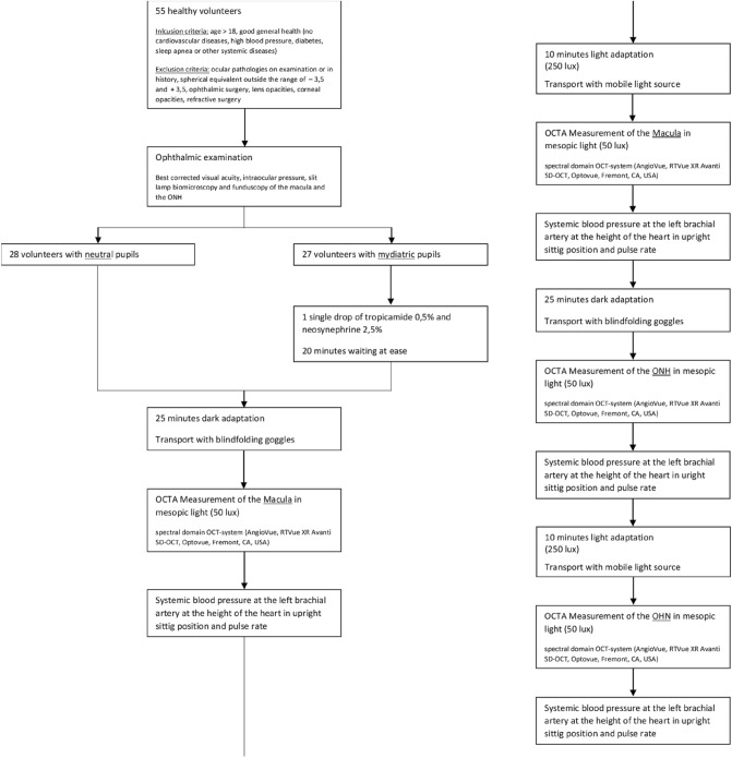
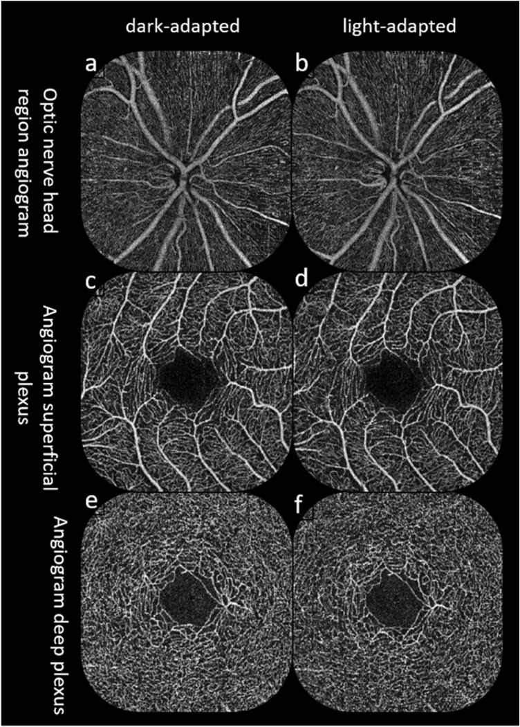
Optical coherence tomography angiography measurements are influenced by a range of environmental factors as blood pressure and physical fitness. The present study aimed to evaluate the effects of light and dark exposure in eyes with neutral and mydriatic pupils on vessel density in the macular and optic nerve head regions, as measured using optical coherence tomography angiography (OCTA). 55 eyes of 55 healthy volunteers (28 patients with neutral pupils; 27.18 ± 4.33 years) were examined using a high-speed and high-resolution spectral-domain OCT XR Avanti system with a split-spectrum amplitude de-correlation angiography algorithm. OCTA imaging was performed after dark adaptation and after exposure to light. The vessel density data of the superficial and deep retinal macular and optic nerve head region OCT-angiogram were analyzed for these two light conditions. Through Bonferroni correction for multiple testing, the p- value was adapted from 0.05 to 0.017. In eyes with neutral pupils, a significant increase was found in the capillary region of the optic nerve head region (p = 0.002), comparing dark- and light-adaptation. In the macular region of eyes with neutral (p = 0.718) and mydriatic pupils (p = 0.043), no significant differences were observed, as were any in the optic nerve head region of the mydriatic eyes (p = 0.797). This observation suggests that light conditions could be a possible factor influencing OCTA measurements. After dark exposure, vessel density data were significantly different between eyes with neutral and mydriatic pupils (nerve head region: p < 0.0001, superficial macula: p < 0.0001, deep macula: p = 0.0025). These data warn for the effect of mydriatic drops on vessel density measurements.
光学相干断层扫描血管造影测量受多种环境因素的影响,如血压和身体健康。本研究旨在评估在中性和散瞳瞳孔的眼睛中,光和暗适应对黄斑和视神经头区域血管密度的影响,使用光学相干断层扫描血管造影(OCTA)进行测量。 55 只健康志愿者的 55 只眼(28 只中性瞳孔患者;27.18±4.33 岁)使用高速高分辨率光谱域 OCT XR Avanti 系统和分裂光谱幅度去相关血管造影算法进行检查。 OCTA 成像在暗适应后和暴露于光后进行。对这两种光照条件下的深层和浅层视网膜黄斑和视神经头区域 OCT-血管造影的血管密度数据进行了分析。通过对多重测试进行 Bonferroni 校正,p 值从 0.05 调整为 0.017。在中性瞳孔的眼中,视神经头区域的毛细血管区域观察到明显增加(p=0.002),与暗适应相比。在中性(p=0.718)和散瞳(p=0.043)瞳孔的眼睛的黄斑区域,未观察到显著差异,以及散瞳眼的视神经头区域(p=0.797)。这一观察表明,光照条件可能是影响 OCTA 测量的一个因素。在暗适应后,中性和散瞳瞳孔的眼中的血管密度数据存在显著差异(视神经头区域:p<0.0001,浅层黄斑:p<0.0001,深层黄斑:p=0.0025)。这些数据警告散瞳滴剂对血管密度测量的影响。