• 文献检索
  • 文档翻译
  • 深度研究
  • 学术资讯
  • Suppr Zotero 插件Zotero 插件
  • 邀请有礼
  • 套餐&价格
  • 历史记录
应用&插件
Suppr Zotero 插件Zotero 插件浏览器插件Mac 客户端Windows 客户端微信小程序
定价
高级版会员购买积分包购买API积分包
服务
文献检索文档翻译深度研究API 文档MCP 服务
关于我们
关于 Suppr公司介绍联系我们用户协议隐私条款
关注我们

Suppr 超能文献

核心技术专利:CN118964589B侵权必究
粤ICP备2023148730 号-1Suppr @ 2026

文献检索

告别复杂PubMed语法,用中文像聊天一样搜索,搜遍4000万医学文献。AI智能推荐,让科研检索更轻松。

立即免费搜索

文件翻译

保留排版,准确专业,支持PDF/Word/PPT等文件格式,支持 12+语言互译。

免费翻译文档

深度研究

AI帮你快速写综述,25分钟生成高质量综述,智能提取关键信息,辅助科研写作。

立即免费体验

扫频源光学相干断层扫描血管造影术的最新进展

The Latest Updates in Swept-Source Optical Coherence Tomography Angiography.

作者信息

Xia Fan, Hua Rui

机构信息

Department of Ophthalmology, The Fourth People's Hospital of Shenyang, China Medical University, Shenyang 110000, China.

Department of Ophthalmology, First Hospital of China Medical University, Shenyang 110001, China.

出版信息

Diagnostics (Basel). 2023 Dec 25;14(1):47. doi: 10.3390/diagnostics14010047.

DOI:10.3390/diagnostics14010047
PMID:38201356
原文链接:https://pmc.ncbi.nlm.nih.gov/articles/PMC10795713/
Abstract

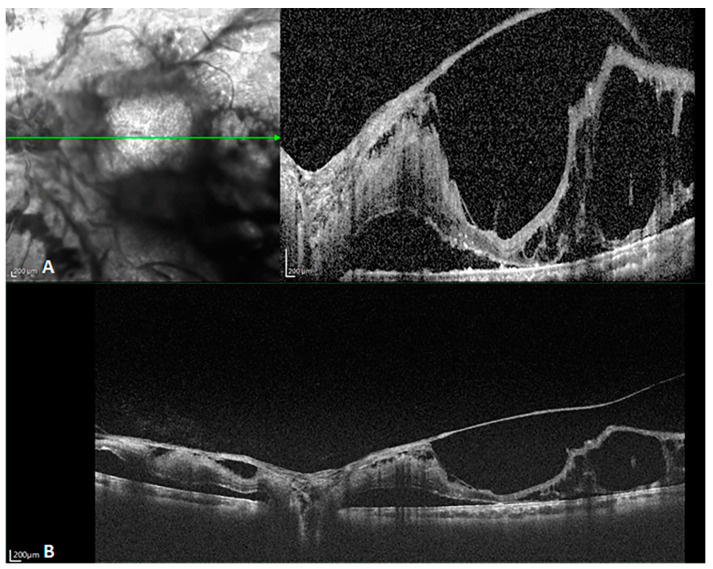
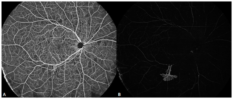
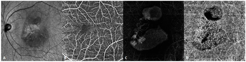
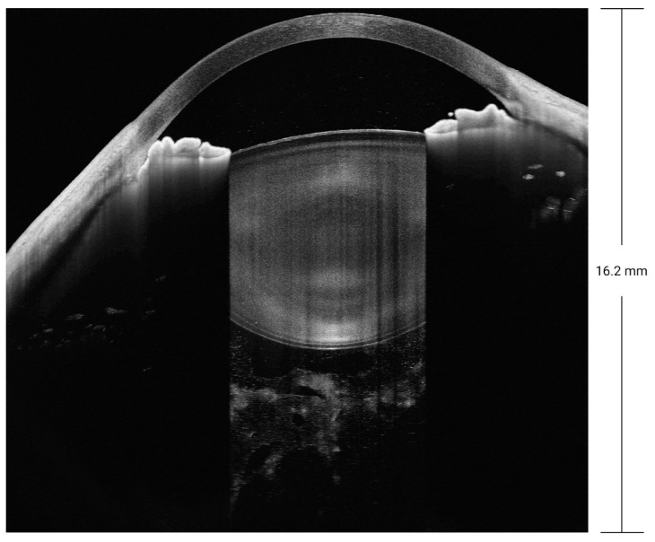
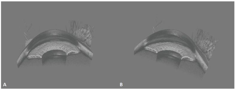
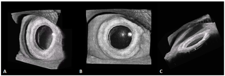
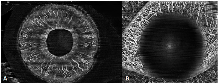
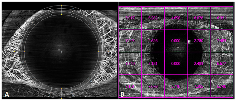
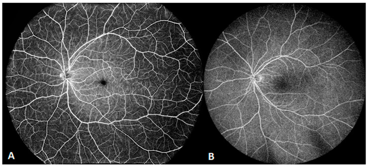
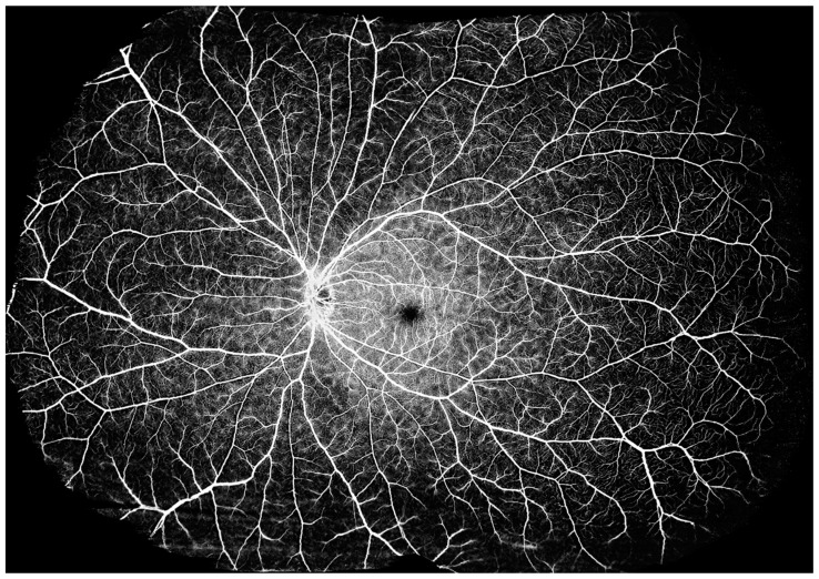
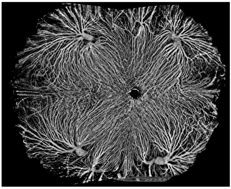
Optical coherence tomography (OCT) is a revolutionary imaging technology in the field of ophthalmic medical imaging [...].

摘要

光学相干断层扫描(OCT)是眼科医学成像领域的一项革命性成像技术[...]。

https://cdn.ncbi.nlm.nih.gov/pmc/blobs/7d70/10795713/d11ca9d2c428/diagnostics-14-00047-g016.jpg
https://cdn.ncbi.nlm.nih.gov/pmc/blobs/7d70/10795713/a0771ef272cb/diagnostics-14-00047-g001.jpg
https://cdn.ncbi.nlm.nih.gov/pmc/blobs/7d70/10795713/3e5b932ef9a2/diagnostics-14-00047-g002.jpg
https://cdn.ncbi.nlm.nih.gov/pmc/blobs/7d70/10795713/e6c4a9770391/diagnostics-14-00047-g003.jpg
https://cdn.ncbi.nlm.nih.gov/pmc/blobs/7d70/10795713/66fa8740fa28/diagnostics-14-00047-g004.jpg
https://cdn.ncbi.nlm.nih.gov/pmc/blobs/7d70/10795713/8d802ee0a30c/diagnostics-14-00047-g005.jpg
https://cdn.ncbi.nlm.nih.gov/pmc/blobs/7d70/10795713/47c7d63a7e9e/diagnostics-14-00047-g006.jpg
https://cdn.ncbi.nlm.nih.gov/pmc/blobs/7d70/10795713/46c1aef43f46/diagnostics-14-00047-g007.jpg
https://cdn.ncbi.nlm.nih.gov/pmc/blobs/7d70/10795713/72df91ef6e55/diagnostics-14-00047-g008.jpg
https://cdn.ncbi.nlm.nih.gov/pmc/blobs/7d70/10795713/cb726dd2bfbf/diagnostics-14-00047-g009.jpg
https://cdn.ncbi.nlm.nih.gov/pmc/blobs/7d70/10795713/5a64ebd21a70/diagnostics-14-00047-g010.jpg
https://cdn.ncbi.nlm.nih.gov/pmc/blobs/7d70/10795713/f5967001515c/diagnostics-14-00047-g011.jpg
https://cdn.ncbi.nlm.nih.gov/pmc/blobs/7d70/10795713/9970e79fefc4/diagnostics-14-00047-g012.jpg
https://cdn.ncbi.nlm.nih.gov/pmc/blobs/7d70/10795713/890496a75adf/diagnostics-14-00047-g013.jpg
https://cdn.ncbi.nlm.nih.gov/pmc/blobs/7d70/10795713/e42146c3090f/diagnostics-14-00047-g014.jpg
https://cdn.ncbi.nlm.nih.gov/pmc/blobs/7d70/10795713/097ae160d5d6/diagnostics-14-00047-g015.jpg
https://cdn.ncbi.nlm.nih.gov/pmc/blobs/7d70/10795713/d11ca9d2c428/diagnostics-14-00047-g016.jpg

相似文献

1
The Latest Updates in Swept-Source Optical Coherence Tomography Angiography.扫频源光学相干断层扫描血管造影术的最新进展
Diagnostics (Basel). 2023 Dec 25;14(1):47. doi: 10.3390/diagnostics14010047.
2
Retinal applications of swept source optical coherence tomography (OCT) and optical coherence tomography angiography (OCTA).扫频源光学相干断层扫描(OCT)和光相干断层扫描血管造影(OCTA)在视网膜中的应用。
Prog Retin Eye Res. 2021 Sep;84:100951. doi: 10.1016/j.preteyeres.2021.100951. Epub 2021 Jan 28.
3
Advances in swept-source optical coherence tomography and optical coherence tomography angiography.扫频源光学相干断层扫描及光学相干断层扫描血管造影术的进展。
Adv Ophthalmol Pract Res. 2022 Nov 25;3(2):67-79. doi: 10.1016/j.aopr.2022.10.005. eCollection 2023 May-Jun.
4
SPECTRAL DOMAIN VERSUS SWEPT SOURCE OPTICAL COHERENCE TOMOGRAPHY ANGIOGRAPHY OF THE RETINAL CAPILLARY PLEXUSES IN SICKLE CELL MACULOPATHY.镰状细胞性黄斑病变中视网膜毛细血管丛的光谱域与扫频源光学相干断层扫描血管造影术
Retin Cases Brief Rep. 2018;12(2):87-92. doi: 10.1097/ICB.0000000000000448.
5
Swept-source optical coherence tomography and optical coherence tomography angiography in acquired toxoplasmic chorioretinitis: a case report.扫频源光学相干断层扫描和光学相干断层扫描血管造影在获得性弓形体性脉络膜视网膜炎中的应用:病例报告
J Med Case Rep. 2018 Dec 4;12(1):358. doi: 10.1186/s13256-018-1902-x.
6
[Swept source optical coherence tomography: a technology review].[扫频光源光学相干断层扫描技术综述]
Vestn Oftalmol. 2020;136(1):111-116. doi: 10.17116/oftalma2020136011111.
7
Clinical utility of anterior segment swept-source optical coherence tomography in glaucoma.眼前节扫频光学相干断层扫描在青光眼诊疗中的临床应用价值
Oman J Ophthalmol. 2016 Jan-Apr;9(1):3-10. doi: 10.4103/0974-620X.176093.
8
Multimodal imaging of hypertensive chorioretinopathy by swept-source optical coherence tomography and optical coherence tomography angiography: Case report.扫频源光学相干断层扫描和光学相干断层扫描血管造影对高血压性脉络膜视网膜病变的多模态成像:病例报告
Medicine (Baltimore). 2017 Sep;96(39):e8110. doi: 10.1097/MD.0000000000008110.
9
Imaging choroidal neovascular membrane using en face swept-source optical coherence tomography angiography.使用正面扫频源光学相干断层扫描血管造影术对脉络膜新生血管膜进行成像。
Clin Ophthalmol. 2017 Oct 13;11:1859-1869. doi: 10.2147/OPTH.S143018. eCollection 2017.
10
Choroidal analysis in healthy eyes using swept-source optical coherence tomography compared to spectral domain optical coherence tomography.应用扫频源光学相干断层扫描与频域光学相干断层扫描对健康眼的脉络膜进行分析。
Am J Ophthalmol. 2014 Jun;157(6):1272-1281.e1. doi: 10.1016/j.ajo.2014.02.034. Epub 2014 Feb 18.

引用本文的文献

1
A Novel Method for Analysing the Curvature of the Anterior Lens: Multi-Radial Scheimpflug Imaging and Custom Conic Fitting Algorithm.一种分析晶状体前表面曲率的新方法:多径向Scheimpflug成像与定制圆锥拟合算法
J Imaging. 2025 Aug 1;11(8):257. doi: 10.3390/jimaging11080257.
2
A novel stress sensor enables accurate estimation of micro-scale tissue mechanics in quantitative micro-elastography.一种新型应力传感器能够在定量微弹性成像中准确估计微观尺度的组织力学。
APL Bioeng. 2024 Sep 23;8(3):036115. doi: 10.1063/5.0220309. eCollection 2024 Sep.
3
Advances in Structural and Functional Retinal Imaging and Biomarkers for Early Detection of Diabetic Retinopathy.

本文引用的文献

1
Light conditions influence optic nerve OCT angiography parameter in healthy subjects with neutral pupils.光照条件会影响瞳孔居中的健康受试者的视神经 OCT 血管造影参数。
Sci Rep. 2023 Jun 6;13(1):9154. doi: 10.1038/s41598-023-36069-5.
2
Thermally tuned VCSEL at 850 nm as a low-cost alternative source for full-eye SS-OCT.850nm 热调谐 VCSEL 作为全眼 SS-OCT 的低成本替代光源。
Opt Lett. 2023 Jun 1;48(11):3079-3082. doi: 10.1364/OL.489050.
3
Optical Coherence Tomography and Optical Coherence Tomography Angiography in Neglected Diseases.
用于糖尿病视网膜病变早期检测的视网膜结构和功能成像及生物标志物的进展
Biomedicines. 2024 Jun 25;12(7):1405. doi: 10.3390/biomedicines12071405.
4
Non-mechanical steering of the optical beam in spectral-domain optical coherence tomography.光谱域光学相干断层扫描中光束的非机械转向
Sci Rep. 2024 Jun 27;14(1):14860. doi: 10.1038/s41598-024-65125-x.
光学相干断层扫描和光学相干断层血管造影在被忽视疾病中的应用。
Ocul Immunol Inflamm. 2024 Sep;32(7):1427-1434. doi: 10.1080/09273948.2023.2211161. Epub 2023 May 19.
4
The Role of Optical Coherence Tomography Angiography in Optic Nerve Head Edema: A Narrative Review.光学相干断层扫描血管造影在视神经乳头水肿中的作用:一项叙述性综述
J Ophthalmol. 2022 Nov 30;2022:5823345. doi: 10.1155/2022/5823345. eCollection 2022.
5
High speed, long range, deep penetration swept source OCT for structural and angiographic imaging of the anterior eye.高速、长程、深穿透扫频源 OCT 用于眼前节的结构和血管成像。
Sci Rep. 2022 Jan 19;12(1):992. doi: 10.1038/s41598-022-04784-0.
6
Optical Coherence Tomography and Glaucoma.光学相干断层扫描与青光眼。
Annu Rev Vis Sci. 2021 Sep 15;7:693-726. doi: 10.1146/annurev-vision-100419-111350. Epub 2021 Jul 9.
7
Retinal applications of swept source optical coherence tomography (OCT) and optical coherence tomography angiography (OCTA).扫频源光学相干断层扫描(OCT)和光相干断层扫描血管造影(OCTA)在视网膜中的应用。
Prog Retin Eye Res. 2021 Sep;84:100951. doi: 10.1016/j.preteyeres.2021.100951. Epub 2021 Jan 28.
8
Anterior segment optical coherence tomography angiography for iris vasculature in pigmented eyes.眼前节光学相干断层扫描血管造影在色素眼虹膜血管中的应用。
Br J Ophthalmol. 2021 Jul;105(7):929-934. doi: 10.1136/bjophthalmol-2020-316930. Epub 2020 Aug 18.