Cui Yuanyuan, Dang Yixuan, Zhang Hao, Peng Hong, Zhang Jun, Li Jinhang, Shen Peiyi, Mao Cuiping, Ma Lin, Zhang Liang
Department of Radiology, The First Medical Center, Chinese PLA General Hospital, Beijing, China.
Department of Radiology, Qingdao Special Servicemen Recuperation Center of PLA Navy, Qingdao, China.
Quant Imaging Med Surg. 2023 Jun 1;13(6):3400-3415. doi: 10.21037/qims-22-887. Epub 2023 May 15.
The present study aimed to establish a robust predictive model based on a machine learning (ML) algorithm providing preoperative noninvasive diagnosis and to further explore the contribution of each magnetic resonance imaging (MRI) sequence to the classification to help select images for future model development.
This was a retrospective cross-sectional study, and consecutive patients with histologically confirmed diffuse gliomas in our hospital from November 2015 to October 2019 were recruited. The participants were grouped into a training and testing set based on a ratio of 8:2. Five MRI sequences were employed to develop the support vector machine (SVM) classification model. An advanced contrast analysis of single-sequence-based classifiers was performed, according to which different sequence combinations were tested, and the best one was selected to form an ultimate classifier. Patients whose MRIs were acquired with other types of scanners formed an additional, independent validation set.
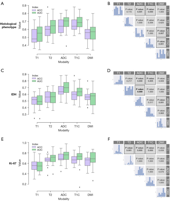
A total of 150 patients with gliomas were used in the present study. Contrast analysis revealed that the contribution of the apparent diffusion coefficient (ADC) was the most significant [accuracies were as follows: histological phenotype, 0.640; isocitrate dehydrogenase (IDH) status, 0.656; and Ki-67 expression, 0.699] and that of T1 weighted imaging was limited (accuracies were as follows: histological phenotype, 0.521; IDH status, 0.492; and Ki-67 expression, 0.556). The ultimate classifiers for IDH status, histological phenotype, and Ki-67 expression achieved promising performances with area under the curve (AUC) values of 0.88, 0.93, and 0.93, respectively. The classifiers for the histological phenotype, IDH status, and Ki-67 expression correctly predicted 3 of 5 subjects, 6 of 7 subjects, and 9 of 13 subjects in the additional validation set, respectively.
The present study showed satisfactory performance in predicting the IDH genotype, histological phenotype, and Ki-67 expression level. The contrast analysis revealed the contribution of different MRI sequences and suggested that the combination of all the acquired sequences was not the optimal strategy to build the radiogenomics-based classifier.
本研究旨在基于机器学习(ML)算法建立一个强大的预测模型,以提供术前无创诊断,并进一步探索每个磁共振成像(MRI)序列对分类的贡献,以帮助选择图像用于未来模型开发。
这是一项回顾性横断面研究,招募了2015年11月至2019年10月在我院组织学确诊为弥漫性胶质瘤的连续患者。参与者按8:2的比例分为训练集和测试集。采用五个MRI序列开发支持向量机(SVM)分类模型。对基于单序列的分类器进行了高级对比分析,据此测试了不同的序列组合,并选择最佳组合形成最终分类器。使用其他类型扫描仪进行MRI检查的患者组成了一个额外的独立验证集。
本研究共纳入150例胶质瘤患者。对比分析显示,表观扩散系数(ADC)的贡献最为显著[准确率如下:组织学表型为0.640;异柠檬酸脱氢酶(IDH)状态为0.656;Ki-67表达为0.699],而T1加权成像的贡献有限(准确率如下:组织学表型为0.521;IDH状态为0.492;Ki-67表达为0.556)。IDH状态、组织学表型和Ki-67表达的最终分类器表现良好,曲线下面积(AUC)值分别为0.88、0.93和0.93。组织学表型、IDH状态和Ki-67表达的分类器在额外验证集中分别正确预测了5名受试者中的3名、7名受试者中的6名和13名受试者中的9名。
本研究在预测IDH基因型、组织学表型和Ki-67表达水平方面表现令人满意。对比分析揭示了不同MRI序列的贡献,并表明获取的所有序列的组合并非构建基于放射基因组学的分类器的最佳策略。