• 文献检索
  • 文档翻译
  • 深度研究
  • 学术资讯
  • Suppr Zotero 插件Zotero 插件
  • 邀请有礼
  • 套餐&价格
  • 历史记录
应用&插件
Suppr Zotero 插件Zotero 插件浏览器插件Mac 客户端Windows 客户端微信小程序
定价
高级版会员购买积分包购买API积分包
服务
文献检索文档翻译深度研究API 文档MCP 服务
关于我们
关于 Suppr公司介绍联系我们用户协议隐私条款
关注我们

Suppr 超能文献

核心技术专利:CN118964589B侵权必究
粤ICP备2023148730 号-1Suppr @ 2026

文献检索

告别复杂PubMed语法,用中文像聊天一样搜索,搜遍4000万医学文献。AI智能推荐,让科研检索更轻松。

立即免费搜索

文件翻译

保留排版,准确专业,支持PDF/Word/PPT等文件格式,支持 12+语言互译。

免费翻译文档

深度研究

AI帮你快速写综述,25分钟生成高质量综述,智能提取关键信息,辅助科研写作。

立即免费体验

多核钠、弥散和灌注 MRI 用于人类脑胶质瘤。

Multi-nuclear sodium, diffusion, and perfusion MRI in human gliomas.

机构信息

UCLA Brain Tumor Imaging Laboratory (BTIL), Center for Computer Vision and Imaging Biomarkers, University of California, Los Angeles, Los Angeles, CA, USA.

Department of Radiological Sciences, David Geffen School of Medicine, University of California, Los Angeles, Los Angeles, CA, USA.

出版信息

J Neurooncol. 2023 Jun;163(2):417-427. doi: 10.1007/s11060-023-04363-x. Epub 2023 Jun 9.

DOI:10.1007/s11060-023-04363-x
PMID:37294422
原文链接:https://pmc.ncbi.nlm.nih.gov/articles/PMC10322966/
Abstract

PURPOSE

There is limited knowledge about the associations between sodium and proton MRI measurements in brain tumors. The purpose of this study was to quantify intra- and intertumoral correlations between sodium, diffusion, and perfusion MRI in human gliomas.

METHODS

Twenty glioma patients were prospectively studied on a 3T MRI system with multinuclear capabilities. Three mutually exclusive tumor volumes of interest (VOIs) were segmented: contrast-enhancing tumor (CET), T2/FLAIR hyperintense non-enhancing tumor (NET), and necrosis. Median and voxel-wise associations between apparent diffusion coefficient (ADC), normalized relative cerebral blood volume (nrCBV), and normalized sodium measurements were quantified for each VOI.

RESULTS

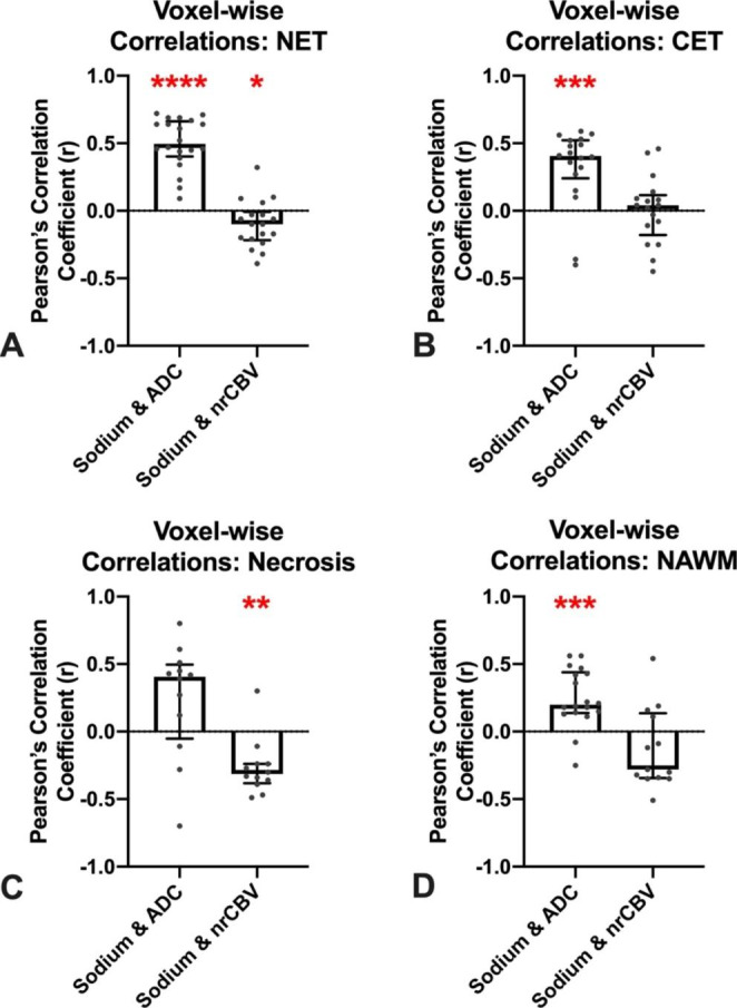
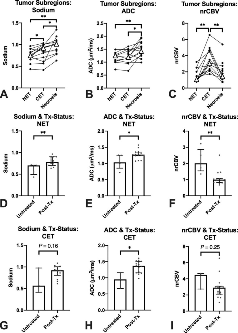
Both relative sodium concentration and ADC were significantly higher in areas of necrosis compared to NET (P = 0.003 and P = 0.008, respectively) and CET (P = 0.02 and P = 0.02). Sodium concentration was higher in CET compared to NET (P = 0.04). Sodium and ADC were higher in treated compared to treatment-naïve gliomas within NET (P = 0.006 and P = 0.01, respectively), and ADC was elevated in CET (P = 0.03). Median ADC and sodium concentration were positively correlated across patients in NET (r = 0.77, P < 0.0001) and CET (r = 0.84, P < 0.0001), but not in areas of necrosis (r = 0.45, P = 0.12). Median nrCBV and sodium concentration were negatively correlated across patients in areas of NET (r=-0.63, P = 0.003). Similar associations were observed when examining voxel-wise correlations within VOIs.

CONCLUSION

Sodium MRI is positively correlated with proton diffusion MRI measurements in gliomas, likely reflecting extracellular water. Unique areas of multinuclear MRI contrast may be useful in future studies to understand the chemistry of the tumor microenvironment.

摘要

目的

目前对于脑肿瘤中钠和质子 MRI 测量之间的相关性知之甚少。本研究的目的是定量研究人类脑胶质瘤中钠、弥散和灌注 MRI 之间的瘤内和瘤间相关性。

方法

20 例胶质瘤患者前瞻性地在具有多核功能的 3T MRI 系统上进行研究。三个相互排斥的肿瘤感兴趣区(VOI)被分割:对比增强肿瘤(CET)、T2/FLAIR 高信号非增强肿瘤(NET)和坏死区。对每个 VOI 量化平均扩散系数(ADC)、归一化相对脑血容量(nrCBV)和归一化钠测量值之间的中位数和体素相关性。

结果

与 NET(P=0.003 和 P=0.008)和 CET(P=0.02 和 P=0.02)相比,坏死区的相对钠浓度和 ADC 均显著升高,与 CET 相比,NET 区的钠浓度更高(P=0.04)。与 NET 内治疗前胶质瘤相比,治疗后胶质瘤的钠浓度和 ADC 更高(P=0.006 和 P=0.01),CET 区的 ADC 升高(P=0.03)。NET(r=0.77,P<0.0001)和 CET(r=0.84,P<0.0001)中,患者间的 ADC 和钠浓度中位数呈正相关,但坏死区(r=0.45,P=0.12)无相关性。NET 区患者的中位 nrCBV 和钠浓度呈负相关(r=-0.63,P=0.003)。在 VOI 内检查体素相关性时也观察到了类似的相关性。

结论

胶质瘤中钠 MRI 与质子弥散 MRI 测量值呈正相关,可能反映细胞外水。多核 MRI 对比的独特区域可能有助于未来研究了解肿瘤微环境的化学性质。

https://cdn.ncbi.nlm.nih.gov/pmc/blobs/7bdd/10322966/87fe85345f0a/11060_2023_4363_Figd_HTML.jpg
https://cdn.ncbi.nlm.nih.gov/pmc/blobs/7bdd/10322966/6d32f8cb5fad/11060_2023_4363_Figa_HTML.jpg
https://cdn.ncbi.nlm.nih.gov/pmc/blobs/7bdd/10322966/7b2010b9d2c2/11060_2023_4363_Figb_HTML.jpg
https://cdn.ncbi.nlm.nih.gov/pmc/blobs/7bdd/10322966/5cf840c21438/11060_2023_4363_Figc_HTML.jpg
https://cdn.ncbi.nlm.nih.gov/pmc/blobs/7bdd/10322966/87fe85345f0a/11060_2023_4363_Figd_HTML.jpg

相似文献

1
Multi-nuclear sodium, diffusion, and perfusion MRI in human gliomas.多核钠、弥散和灌注 MRI 用于人类脑胶质瘤。
J Neurooncol. 2023 Jun;163(2):417-427. doi: 10.1007/s11060-023-04363-x. Epub 2023 Jun 9.
2
Diffusion MRI is superior to quantitative T2-FLAIR mismatch in predicting molecular subtypes of human non-enhancing gliomas.在预测人类非强化型胶质瘤的分子亚型方面,扩散加权磁共振成像优于定量T2液体衰减反转恢复序列不匹配。
Neuroradiology. 2024 Dec;66(12):2153-2162. doi: 10.1007/s00234-024-03475-z. Epub 2024 Oct 8.
3
Magnetic resonance perfusion for differentiating low-grade from high-grade gliomas at first presentation.首次就诊时磁共振灌注成像用于鉴别低级别与高级别胶质瘤
Cochrane Database Syst Rev. 2018 Jan 22;1(1):CD011551. doi: 10.1002/14651858.CD011551.pub2.
4
Supratentorial glioma grading in children by using apparent diffusion coefficient map: application of histogram analysis based on segmentation.基于分割的直方图分析在儿童幕上胶质瘤表观扩散系数图分级中的应用
Clin Ter. 2025;176(3):301-309. doi: 10.7417/CT.2025.5226.
5
Simultaneous quantification of perfusion, permeability, and leakage effects in brain gliomas using dynamic spin-and-gradient-echo echoplanar imaging MRI.使用动态自旋梯度回波平面成像 MRI 同时定量脑胶质瘤中的灌注、渗透性和漏出效应。
Eur Radiol. 2024 May;34(5):3087-3101. doi: 10.1007/s00330-023-10215-z. Epub 2023 Oct 26.
6
Super-resolution sodium MRI of human gliomas at 3T using physics-based generative artificial intelligence.使用基于物理的生成式人工智能在3T条件下对人脑胶质瘤进行超分辨率钠磁共振成像。
J Neurooncol. 2025 Jun 3. doi: 10.1007/s11060-025-05094-x.
7
Pseudo-Resting-State Functional MRI Derived from Dynamic Susceptibility Contrast Perfusion MRI Can Predict Cognitive Impairment in Glioma.动态磁敏感对比灌注 MRI 衍生的假性静息态功能 MRI 可预测脑肿瘤患者认知障碍。
AJNR Am J Neuroradiol. 2024 Oct 3;45(10):1552-1561. doi: 10.3174/ajnr.A8327.
8
Imaging-Based Molecular Characterization of Adult-Type Diffuse Glioma Using Diffusion and Perfusion MRI in Pre- and Post-Treatment Stage Considering Spatial and Temporal Heterogeneity.考虑空间和时间异质性,在治疗前和治疗后阶段使用扩散加权磁共振成像(DWI)和灌注加权磁共振成像(PWI)对成人型弥漫性胶质瘤进行基于影像的分子特征分析
J Magn Reson Imaging. 2025 Aug;62(2):468-479. doi: 10.1002/jmri.29781. Epub 2025 Apr 8.
9
Perfusion Showdown: Comparison of Multiple MRI Perfusion Techniques in the Grading of Pediatric Brain Tumors.灌注对决:多种MRI灌注技术在小儿脑肿瘤分级中的比较
AJNR Am J Neuroradiol. 2024 Dec 23. doi: 10.3174/ajnr.A8635.
10
Deep learning based apparent diffusion coefficient map generation from multi-parametric MR images for patients with diffuse gliomas.基于深度学习从多参数磁共振图像生成弥漫性胶质瘤患者的表观扩散系数图。
Med Phys. 2025 Feb;52(2):847-855. doi: 10.1002/mp.17509. Epub 2024 Nov 8.

引用本文的文献

1
Gray matter sodium accumulation and atrophy in relapsing-remitting multiple sclerosis correspond to neurotransmitter maps.复发缓解型多发性硬化症中的灰质钠积累和萎缩与神经递质图谱相对应。
Eur Radiol. 2025 Jul 31. doi: 10.1007/s00330-025-11892-8.
2
Super-resolution sodium MRI of human gliomas at 3T using physics-based generative artificial intelligence.使用基于物理的生成式人工智能在3T条件下对人脑胶质瘤进行超分辨率钠磁共振成像。
J Neurooncol. 2025 Jun 3. doi: 10.1007/s11060-025-05094-x.
3
Multinuclear Interleaving of H CEST, Water T*, and Na MRI at 3 T.

本文引用的文献

1
A multi-reader comparison of normal-appearing white matter normalization techniques for perfusion and diffusion MRI in brain tumors.多阅读者比较正常表现的脑肿瘤灌注和弥散 MRI 的白质归一化技术。
Neuroradiology. 2023 Mar;65(3):559-568. doi: 10.1007/s00234-022-03072-y. Epub 2022 Oct 27.
2
Repeatability of simultaneous 3D H MRF/Na MRI in brain at 7 T.7T 下大脑同步 3D H MRF/Na MRI 的可重复性。
Sci Rep. 2022 Aug 19;12(1):14156. doi: 10.1038/s41598-022-18388-1.
3
Sodium MR Neuroimaging.磁共振神经成像中的钠成像。
3T下H CEST、水T*和钠MRI的多核交织
NMR Biomed. 2025 Mar;38(3):e70003. doi: 10.1002/nbm.70003.
4
Radiosurgery-induced early changes in peritumoral tissue sodium concentration of brain metastases.脑转移瘤瘤周组织钠浓度的放射外科诱导早期变化。
PLoS One. 2024 Nov 4;19(11):e0313199. doi: 10.1371/journal.pone.0313199. eCollection 2024.
5
Standardized brain tumor imaging protocols for clinical trials: current recommendations and tips for integration.用于临床试验的标准化脑肿瘤成像方案:当前建议及整合要点
Front Radiol. 2023 Dec 13;3:1267615. doi: 10.3389/fradi.2023.1267615. eCollection 2023.
6
Diagnostic and Therapeutic Issues in Glioma Using Imaging Data: The Challenge of Numerical Twinning.利用影像数据处理神经胶质瘤的诊断与治疗问题:数字孪生的挑战
J Clin Med. 2023 Dec 15;12(24):7706. doi: 10.3390/jcm12247706.
7
The use of sodium MRI in the diagnosis of an anaplastic astrocytoma during immunotherapy: a case report.钠 MRI 在免疫治疗期间诊断间变性星形细胞瘤中的应用:一例报告。
Childs Nerv Syst. 2024 Mar;40(3):965-967. doi: 10.1007/s00381-023-06195-6. Epub 2023 Oct 25.
AJNR Am J Neuroradiol. 2021 Nov;42(11):1920-1926. doi: 10.3174/ajnr.A7261. Epub 2021 Aug 26.
4
Detection of early changes in the post-radiosurgery vestibular schwannoma microenvironment using multinuclear MRI.采用多核 MRI 检测放射外科治疗后听神经鞘瘤微环境的早期变化。
Sci Rep. 2021 Aug 3;11(1):15712. doi: 10.1038/s41598-021-95022-6.
5
The 2021 WHO Classification of Tumors of the Central Nervous System: a summary.2021 年世卫组织中枢神经系统肿瘤分类:概述。
Neuro Oncol. 2021 Aug 2;23(8):1231-1251. doi: 10.1093/neuonc/noab106.
6
Ultra-high-field sodium MRI as biomarker for tumor extent, grade and IDH mutation status in glioma patients.超高场强钠磁共振成像作为胶质瘤患者肿瘤范围、分级及异柠檬酸脱氢酶(IDH)突变状态的生物标志物
Neuroimage Clin. 2020;28:102427. doi: 10.1016/j.nicl.2020.102427. Epub 2020 Sep 12.
7
Relaxometry and quantification in sodium MRI of cerebral gliomas: A FET-PET and MRI small-scale study.脑胶质瘤钠 MRI 的弛豫率测定和定量:FET-PET 和 MRI 小范围研究。
NMR Biomed. 2020 Oct;33(10):e4361. doi: 10.1002/nbm.4361. Epub 2020 Jul 21.
8
Comparison of [F]Fluoroethyltyrosine PET and Sodium MRI in Cerebral Gliomas: a Pilot Study.氟乙基酪氨酸 PET 与钠 MRI 在脑胶质瘤中的对比:一项初步研究。
Mol Imaging Biol. 2020 Feb;22(1):198-207. doi: 10.1007/s11307-019-01349-y.
9
Validation of vessel size imaging (VSI) in high-grade human gliomas using magnetic resonance imaging, image-guided biopsies, and quantitative immunohistochemistry.采用磁共振成像、影像引导下活检和定量免疫组织化学技术验证高级别人脑胶质瘤中的血管大小成像(VSI)。
Sci Rep. 2019 Feb 26;9(1):2846. doi: 10.1038/s41598-018-37564-w.
10
Residual Tumor Volume, Cell Volume Fraction, and Tumor Cell Kill During Fractionated Chemoradiation Therapy of Human Glioblastoma using Quantitative Sodium MR Imaging.使用定量钠磁共振成像对人胶质母细胞瘤进行分割放化疗期间的残余肿瘤体积、细胞体积分数和肿瘤细胞杀伤。
Clin Cancer Res. 2019 Feb 15;25(4):1226-1232. doi: 10.1158/1078-0432.CCR-18-2079. Epub 2018 Nov 28.