• 文献检索
  • 文档翻译
  • 深度研究
  • 学术资讯
  • Suppr Zotero 插件Zotero 插件
  • 邀请有礼
  • 套餐&价格
  • 历史记录
应用&插件
Suppr Zotero 插件Zotero 插件浏览器插件Mac 客户端Windows 客户端微信小程序
定价
高级版会员购买积分包购买API积分包
服务
文献检索文档翻译深度研究API 文档MCP 服务
关于我们
关于 Suppr公司介绍联系我们用户协议隐私条款
关注我们

Suppr 超能文献

核心技术专利:CN118964589B侵权必究
粤ICP备2023148730 号-1Suppr @ 2026

文献检索

告别复杂PubMed语法,用中文像聊天一样搜索,搜遍4000万医学文献。AI智能推荐,让科研检索更轻松。

立即免费搜索

文件翻译

保留排版,准确专业,支持PDF/Word/PPT等文件格式,支持 12+语言互译。

免费翻译文档

深度研究

AI帮你快速写综述,25分钟生成高质量综述,智能提取关键信息,辅助科研写作。

立即免费体验

电突触耦合解决了动作电位波速与心脏中不和谐的交替空间尺度之间的悖论。

Ephaptic Coupling as a Resolution to the Paradox of Action Potential Wave Speed and Discordant Alternans Spatial Scales in the Heart.

机构信息

Rochester Institute of Technology, Rochester, New York 14623, USA.

Hampden-Sydney College, Hampden-Sydney, Virginia 23943, USA.

出版信息

Phys Rev Lett. 2023 May 26;130(21):218401. doi: 10.1103/PhysRevLett.130.218401.

DOI:10.1103/PhysRevLett.130.218401
PMID:37295103
原文链接:https://pmc.ncbi.nlm.nih.gov/articles/PMC10688031/
Abstract

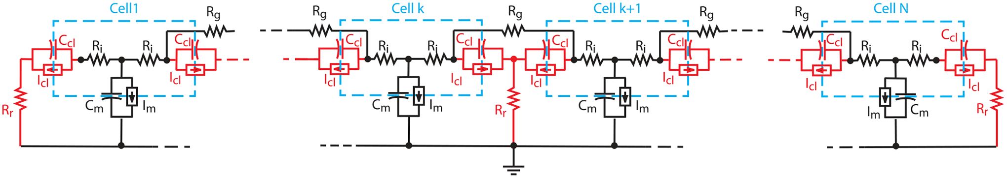
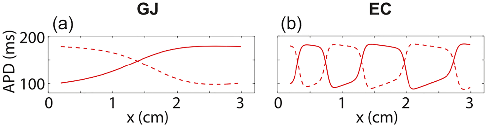
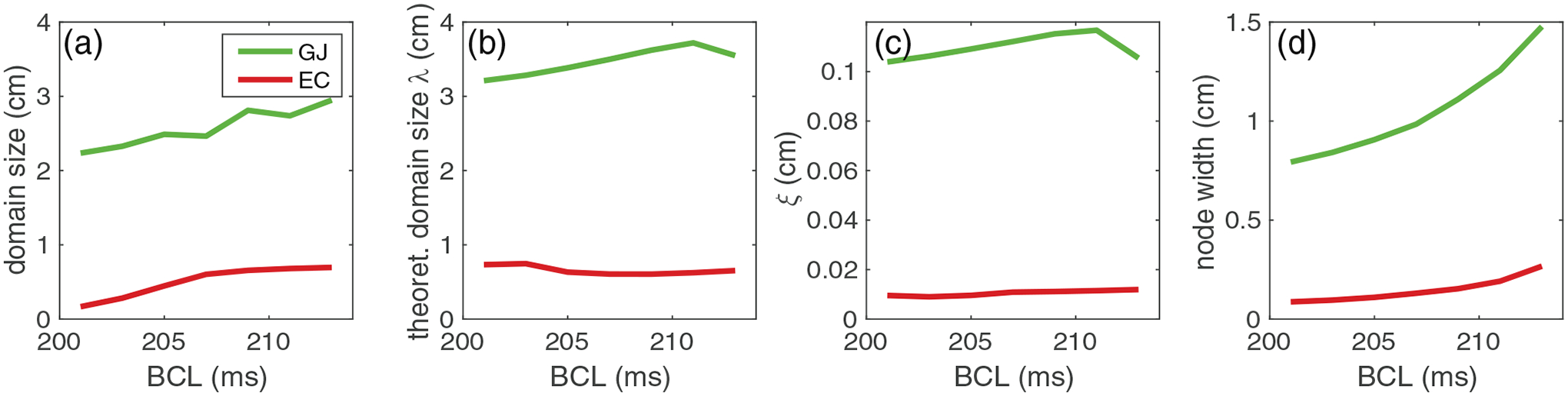
Previous computer simulations have suggested that existing models of action potential wave propagation in the heart are not consistent with observed wave propagation behavior. Specifically, computer models cannot simultaneously reproduce the rapid wave speeds and small spatial scales of discordant alternans patterns measured experimentally in the same simulation. The discrepancy is important, because discordant alternans can be a key precursor to the development of abnormal and dangerous rapid rhythms in the heart. In this Letter, we show that this paradox can be resolved by allowing so-called ephaptic coupling to play a primary role in wave front propagation in place of conventional gap-junction coupling. With this modification, physiological wave speeds and small discordant alternans spatial scales both occur with gap-junction resistance values that are more in line with those observed in experiments. Our theory thus also provides support to the hypothesis that ephaptic coupling plays an important role in normal wave propagation.

摘要

先前的计算机模拟表明,现有的心脏动作电位波传播模型与观察到的波传播行为不一致。具体来说,计算机模型不能在同一模拟中同时再现实验测量到的快速波速和不和谐交替模式的小空间尺度。这种差异很重要,因为不和谐交替可能是心脏异常和危险快速节律发展的关键前兆。在这封信中,我们表明,通过允许所谓的电突触耦合在波前传播中发挥主要作用,而不是传统的缝隙连接耦合,可以解决这个悖论。通过这种修改,生理波速和小的不和谐交替空间尺度都出现在与实验观察到的更一致的缝隙连接电阻值下。因此,我们的理论也支持电突触耦合在正常波传播中起着重要作用的假说。

https://cdn.ncbi.nlm.nih.gov/pmc/blobs/b13f/10688031/d2be46dcd2d7/nihms-1914699-f0004.jpg
https://cdn.ncbi.nlm.nih.gov/pmc/blobs/b13f/10688031/58097907241f/nihms-1914699-f0001.jpg
https://cdn.ncbi.nlm.nih.gov/pmc/blobs/b13f/10688031/7a71c7d9c7d0/nihms-1914699-f0002.jpg
https://cdn.ncbi.nlm.nih.gov/pmc/blobs/b13f/10688031/49205e20e0a9/nihms-1914699-f0003.jpg
https://cdn.ncbi.nlm.nih.gov/pmc/blobs/b13f/10688031/d2be46dcd2d7/nihms-1914699-f0004.jpg

相似文献

1
Ephaptic Coupling as a Resolution to the Paradox of Action Potential Wave Speed and Discordant Alternans Spatial Scales in the Heart.电突触耦合解决了动作电位波速与心脏中不和谐的交替空间尺度之间的悖论。
Phys Rev Lett. 2023 May 26;130(21):218401. doi: 10.1103/PhysRevLett.130.218401.
2
Role of ephaptic coupling in discordant alternans domain sizes and action potential propagation in the heart.电突触耦合在心脏中不和谐的折返波大小和动作电位传播中的作用。
Phys Rev E. 2023 May;107(5-1):054407. doi: 10.1103/PhysRevE.107.054407.
3
Mechanisms for discordant alternans.不协调交替脉的机制。
J Cardiovasc Electrophysiol. 2001 Feb;12(2):196-206. doi: 10.1046/j.1540-8167.2001.00196.x.
4
Ephaptic coupling in cardiac myocytes.心肌细胞的电突触耦合。
IEEE Trans Biomed Eng. 2013 Feb;60(2):576-82. doi: 10.1109/TBME.2012.2226720.
5
Dynamic origin of spatially discordant alternans in cardiac tissue.心脏组织中空间不协调交替现象的动态起源
Biophys J. 2007 Jan 15;92(2):448-60. doi: 10.1529/biophysj.106.091009. Epub 2006 Oct 27.
6
Cardiac alternans induced by fibroblast-myocyte coupling: mechanistic insights from computational models.成纤维细胞-心肌细胞偶联诱导的心脏交替变化:计算模型的机制见解
Am J Physiol Heart Circ Physiol. 2009 Aug;297(2):H775-84. doi: 10.1152/ajpheart.00341.2009. Epub 2009 May 29.
7
Simultaneous Quantification of Spatially Discordant Alternans in Voltage and Intracellular Calcium in Langendorff-Perfused Rabbit Hearts and Inconsistencies with Models of Cardiac Action Potentials and Ca Transients.在Langendorff灌注兔心脏中同时定量电压和细胞内钙的空间不一致交替变化以及与心脏动作电位和钙瞬变模型的不一致性。
Front Physiol. 2017 Oct 20;8:819. doi: 10.3389/fphys.2017.00819. eCollection 2017.
8
Action potential duration dispersion and alternans in simulated heterogeneous cardiac tissue with a structural barrier.具有结构屏障的模拟异质性心脏组织中的动作电位时程离散和交替现象
Biophys J. 2007 Feb 15;92(4):1138-49. doi: 10.1529/biophysj.106.090845. Epub 2006 Nov 17.
9
Constant DI pacing suppresses cardiac alternans formation in numerical cable models.在数值电缆模型中,恒流 DI 起搏可抑制心脏交替现象的形成。
Chaos. 2017 Sep;27(9):093903. doi: 10.1063/1.4999355.
10
Stability of spatially discordant repolarization alternans in cardiac tissue.空间非一致复极交替的稳定性。
Chaos. 2020 Dec;30(12):123141. doi: 10.1063/5.0029209.

引用本文的文献

1
Disruption of ventricular activation by subthreshold delayed afterdepolarizations in RyR2-R420Q catecholaminergic polymorphic ventricular tachycardia.在RyR2-R420Q儿茶酚胺能多形性室性心动过速中,阈下延迟后除极对心室激活的破坏作用
J Mol Cell Cardiol Plus. 2025 Jun 11;13:100466. doi: 10.1016/j.jmccpl.2025.100466. eCollection 2025 Sep.
2
Mechanism of Arrhythmogenesis Driven by Early After Depolarizations in Cardiac Tissue.心脏组织中早期后去极化驱动的心律失常发生机制。
PLoS Comput Biol. 2025 Apr 22;21(4):e1012635. doi: 10.1371/journal.pcbi.1012635. eCollection 2025 Apr.
3
Role of ephaptic coupling in discordant alternans domain sizes and action potential propagation in the heart.

本文引用的文献

1
Role of ephaptic coupling in discordant alternans domain sizes and action potential propagation in the heart.电突触耦合在心脏中不和谐的折返波大小和动作电位传播中的作用。
Phys Rev E. 2023 May;107(5-1):054407. doi: 10.1103/PhysRevE.107.054407.
2
Simulation of atrial fibrillation in a non-ohmic propagation model with dynamic gap junctions.在具有动态缝隙连接的非欧姆传播模型中模拟心房颤动。
Chaos. 2022 Apr;32(4):043113. doi: 10.1063/5.0082763.
3
Cellular Size, Gap Junctions, and Sodium Channel Properties Govern Developmental Changes in Cardiac Conduction.
电突触耦合在心脏中不和谐的折返波大小和动作电位传播中的作用。
Phys Rev E. 2023 May;107(5-1):054407. doi: 10.1103/PhysRevE.107.054407.
细胞大小、缝隙连接和钠通道特性决定心脏传导的发育变化。
Front Physiol. 2021 Oct 25;12:731025. doi: 10.3389/fphys.2021.731025. eCollection 2021.
4
Localization of Na channel clusters in narrowed perinexi of gap junctions enhances cardiac impulse transmission via ephaptic coupling: a model study.缝隙连接变窄处的钠通道簇定位增强了电突触耦合的心脏冲动传递:一项模型研究。
J Physiol. 2021 Nov;599(21):4779-4811. doi: 10.1113/JP282105. Epub 2021 Oct 4.
5
Computational modeling of aberrant electrical activity following remuscularization with intramyocardially injected pluripotent stem cell-derived cardiomyocytes.心肌内注射多能干细胞源性心肌细胞后异常电活动的计算建模。
J Mol Cell Cardiol. 2022 Jan;162:97-109. doi: 10.1016/j.yjmcc.2021.08.011. Epub 2021 Sep 3.
6
Quantifying arrhythmic long QT effects of hydroxychloroquine and azithromycin with whole-heart optical mapping and simulations.利用全心光学映射和模拟量化羟氯喹和阿奇霉素的心律失常性长QT效应。
Heart Rhythm O2. 2021 Jul 3;2(4):394-404. doi: 10.1016/j.hroo.2021.06.008. eCollection 2021 Aug.
7
Intercalated disk nanoscale structure regulates cardiac conduction.闰盘纳米结构调节心脏传导。
J Gen Physiol. 2021 Aug 2;153(8). doi: 10.1085/jgp.202112897. Epub 2021 Jul 15.
8
Mechanisms underlying age-associated manifestation of cardiac sodium channel gain-of-function.心脏钠通道功能获得性与年龄相关表现的潜在机制。
J Mol Cell Cardiol. 2021 Apr;153:60-71. doi: 10.1016/j.yjmcc.2020.12.008. Epub 2020 Dec 26.
9
Intercellular Sodium Regulates Repolarization in Cardiac Tissue with Sodium Channel Gain of Function.细胞间钠离子调控钠离子通道功能获得性心脏组织复极化。
Biophys J. 2020 Jun 2;118(11):2829-2843. doi: 10.1016/j.bpj.2020.04.014. Epub 2020 Apr 21.
10
Spatially Discordant Repolarization Alternans in the Absence of Conduction Velocity Restitution.在无传导速度恢复情况下的空间不协调复极交替
Biophys J. 2020 May 19;118(10):2574-2587. doi: 10.1016/j.bpj.2020.02.008. Epub 2020 Feb 15.