• 文献检索
  • 文档翻译
  • 深度研究
  • 学术资讯
  • Suppr Zotero 插件Zotero 插件
  • 邀请有礼
  • 套餐&价格
  • 历史记录
应用&插件
Suppr Zotero 插件Zotero 插件浏览器插件Mac 客户端Windows 客户端微信小程序
定价
高级版会员购买积分包购买API积分包
服务
文献检索文档翻译深度研究API 文档MCP 服务
关于我们
关于 Suppr公司介绍联系我们用户协议隐私条款
关注我们

Suppr 超能文献

核心技术专利:CN118964589B侵权必究
粤ICP备2023148730 号-1Suppr @ 2026

文献检索

告别复杂PubMed语法,用中文像聊天一样搜索,搜遍4000万医学文献。AI智能推荐,让科研检索更轻松。

立即免费搜索

文件翻译

保留排版,准确专业,支持PDF/Word/PPT等文件格式,支持 12+语言互译。

免费翻译文档

深度研究

AI帮你快速写综述,25分钟生成高质量综述,智能提取关键信息,辅助科研写作。

立即免费体验

闰盘纳米结构调节心脏传导。

Intercalated disk nanoscale structure regulates cardiac conduction.

机构信息

The Ohio State University, Columbus, OH.

Davis Heart and Lung Research Institute, The Ohio State University Wexner Medical Center, Columbus, OH.

出版信息

J Gen Physiol. 2021 Aug 2;153(8). doi: 10.1085/jgp.202112897. Epub 2021 Jul 15.

DOI:10.1085/jgp.202112897
PMID:34264306
原文链接:https://pmc.ncbi.nlm.nih.gov/articles/PMC8287520/
Abstract

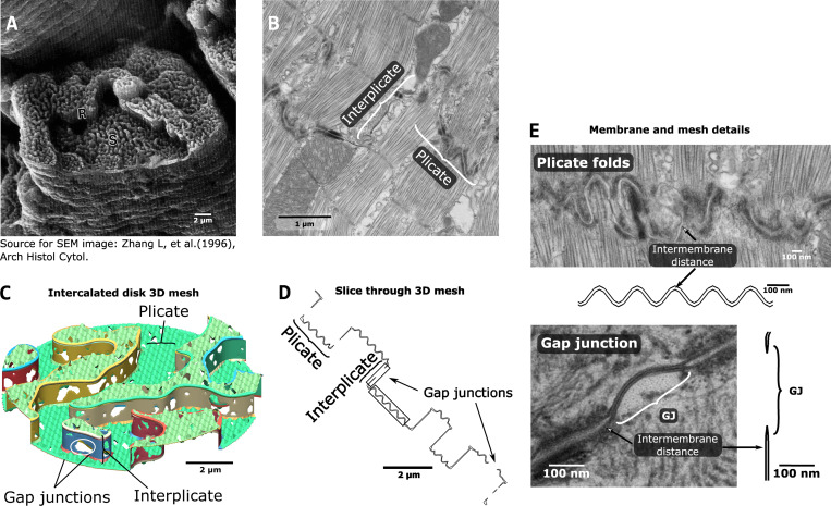
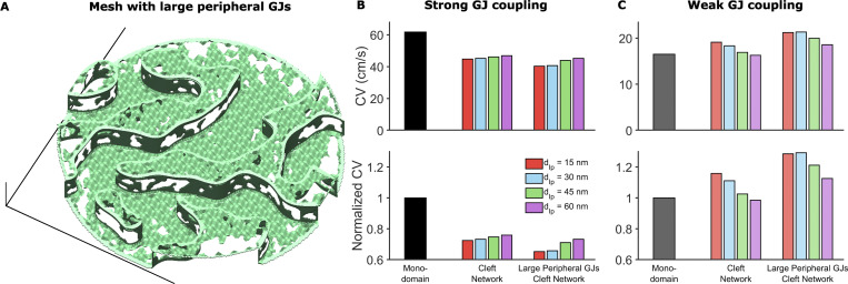
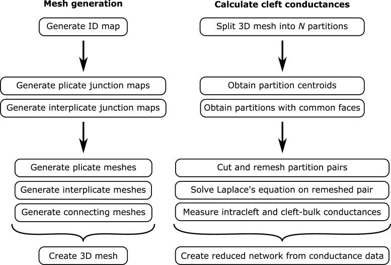
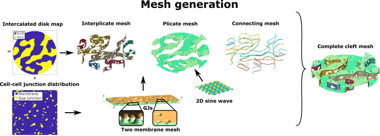
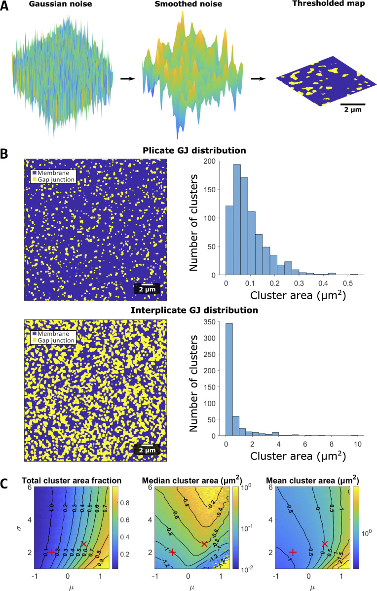
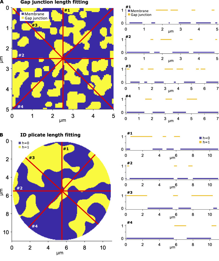
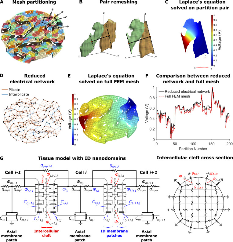
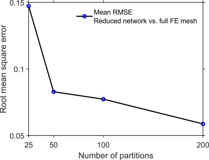
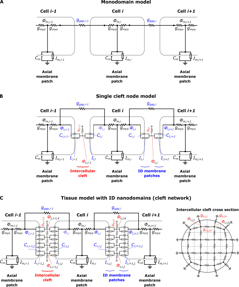
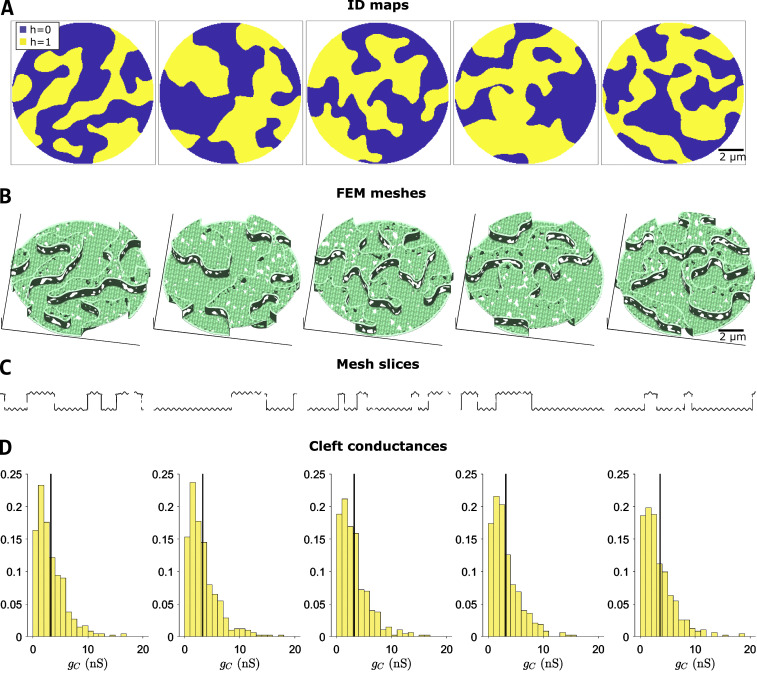
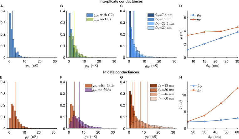
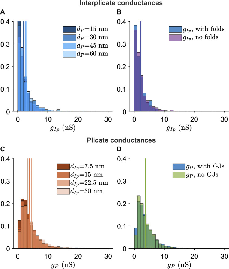
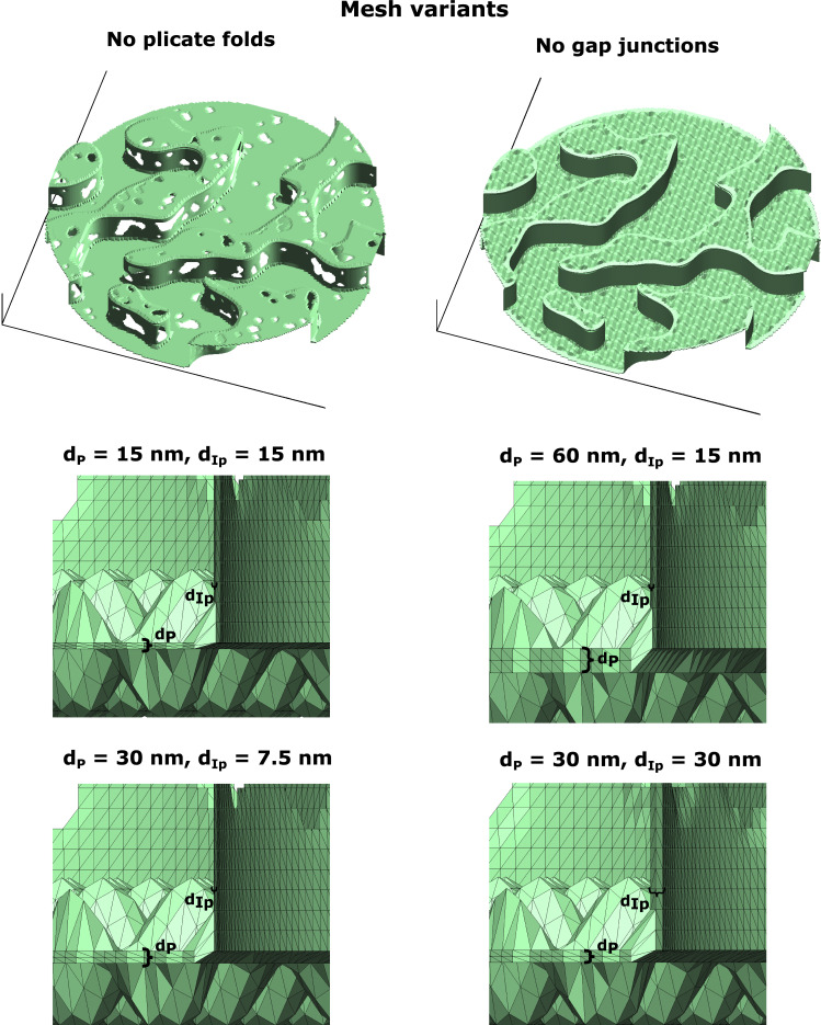
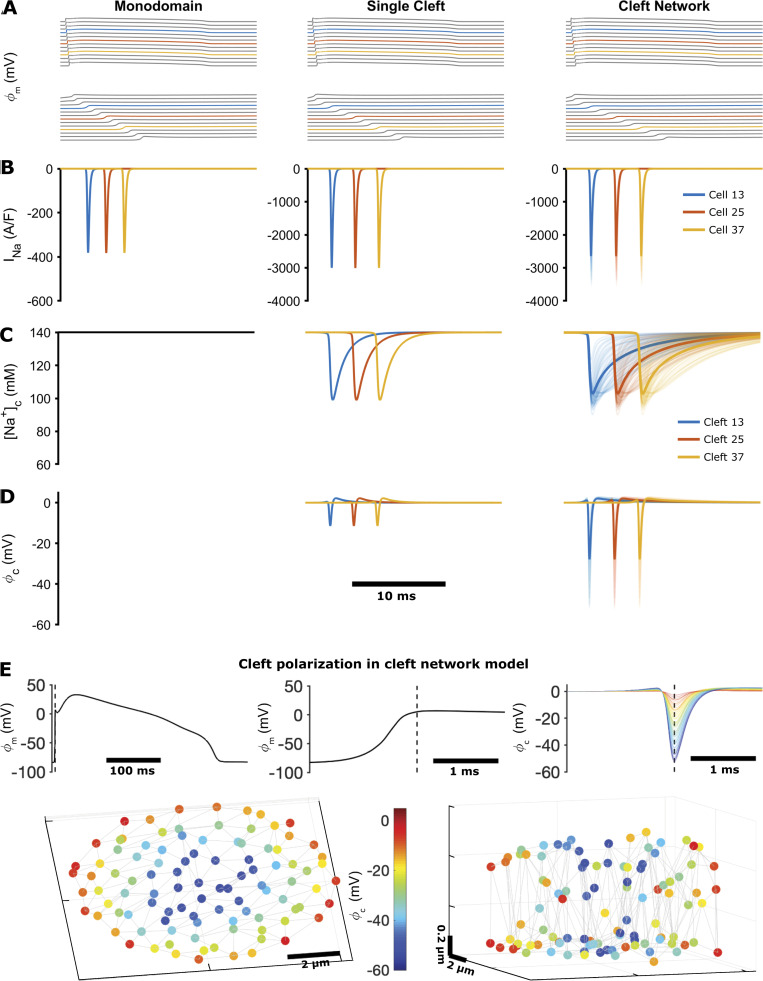
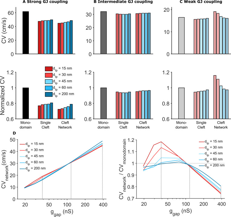
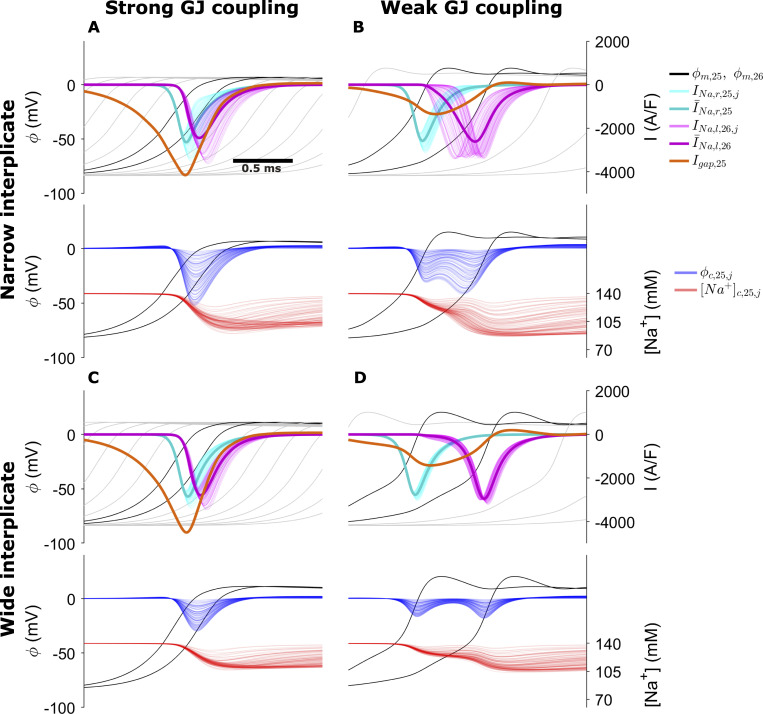
The intercalated disk (ID) is a specialized subcellular region that provides electrical and mechanical connections between myocytes in the heart. The ID has a clearly defined passive role in cardiac tissue, transmitting mechanical forces and electrical currents between cells. Recent studies have shown that Na+ channels, the primary current responsible for cardiac excitation, are preferentially localized at the ID, particularly within nanodomains such as the gap junction-adjacent perinexus and mechanical junction-associated adhesion-excitability nodes, and that perturbations of ID structure alter cardiac conduction. This suggests that the ID may play an important, active role in regulating conduction. However, the structures of the ID and intercellular cleft are not well characterized and, to date, no models have incorporated the influence of ID structure on conduction in cardiac tissue. In this study, we developed an approach to generate realistic finite element model (FEM) meshes replicating nanoscale of the ID structure, based on experimental measurements from transmission electron microscopy images. We then integrated measurements of the intercellular cleft electrical conductivity, derived from the FEM meshes, into a novel cardiac tissue model formulation. FEM-based calculations predict that the distribution of cleft conductances is sensitive to regional changes in ID structure, specifically the intermembrane separation and gap junction distribution. Tissue-scale simulations predict that ID structural heterogeneity leads to significant spatial variation in electrical polarization within the intercellular cleft. Importantly, we found that this heterogeneous cleft polarization regulates conduction by desynchronizing the activation of postjunctional Na+ currents. Additionally, these heterogeneities lead to a weaker dependence of conduction velocity on gap junctional coupling, compared with prior modeling formulations that neglect or simplify ID structure. Further, we found that disruption of local ID nanodomains can either slow or enhance conduction, depending on gap junctional coupling strength. Our study therefore suggests that ID nanoscale structure can play a significant role in regulating cardiac conduction.

摘要

闰盘(ID)是一种特化的亚细胞区域,为心脏中的心肌细胞提供电和机械连接。ID 在心脏组织中具有明确的被动作用,在细胞之间传递机械力和电流。最近的研究表明,Na+通道,即负责心脏兴奋的主要电流,优先定位于 ID,特别是在间隙连接相邻的周质、机械连接相关的粘着-兴奋性节点等纳米域内,并且 ID 结构的扰动会改变心脏传导。这表明 ID 可能在调节传导中发挥重要的主动作用。然而,ID 的结构和细胞间缝隙的结构尚未很好地表征,迄今为止,没有模型将 ID 结构对心脏组织传导的影响纳入其中。在这项研究中,我们开发了一种方法,基于透射电子显微镜图像的实验测量,生成复制 ID 结构纳米尺度的真实有限元模型(FEM)网格。然后,我们将从 FEM 网格中得出的细胞间缝隙电导率的测量值整合到一个新的心脏组织模型公式中。基于 FEM 的计算预测,缝隙电导的分布对 ID 结构的区域变化敏感,特别是膜间分离和缝隙连接的分布。组织尺度的模拟预测,ID 结构异质性导致细胞间缝隙内的电极化存在显著的空间变化。重要的是,我们发现这种异质的缝隙极化通过使后突触 Na+电流的激活不同步来调节传导。此外,与忽略或简化 ID 结构的先前建模公式相比,这些异质性导致传导速度对缝隙连接偶联的依赖性减弱。此外,我们发现局部 ID 纳米域的破坏可能会减缓或增强传导,具体取决于缝隙连接偶联的强度。因此,我们的研究表明 ID 纳米尺度结构可以在调节心脏传导中发挥重要作用。

https://cdn.ncbi.nlm.nih.gov/pmc/blobs/6d35/8287520/053ae48d823d/JGP_202112897_FigS9.jpg
https://cdn.ncbi.nlm.nih.gov/pmc/blobs/6d35/8287520/7202a456b59d/JGP_202112897_Fig1.jpg
https://cdn.ncbi.nlm.nih.gov/pmc/blobs/6d35/8287520/0abe1b2e8e28/JGP_202112897_FigS1.jpg
https://cdn.ncbi.nlm.nih.gov/pmc/blobs/6d35/8287520/97b82432f329/JGP_202112897_Fig2.jpg
https://cdn.ncbi.nlm.nih.gov/pmc/blobs/6d35/8287520/3d5d69440117/JGP_202112897_FigS2.jpg
https://cdn.ncbi.nlm.nih.gov/pmc/blobs/6d35/8287520/c8b2572e7346/JGP_202112897_FigS3.jpg
https://cdn.ncbi.nlm.nih.gov/pmc/blobs/6d35/8287520/3d080890f0dd/JGP_202112897_Fig3.jpg
https://cdn.ncbi.nlm.nih.gov/pmc/blobs/6d35/8287520/adec8575fbce/JGP_202112897_FigS4.jpg
https://cdn.ncbi.nlm.nih.gov/pmc/blobs/6d35/8287520/2ae799585f95/JGP_202112897_FigS5.jpg
https://cdn.ncbi.nlm.nih.gov/pmc/blobs/6d35/8287520/605df06bb2b3/JGP_202112897_FigS6.jpg
https://cdn.ncbi.nlm.nih.gov/pmc/blobs/6d35/8287520/800607db3c24/JGP_202112897_Fig4.jpg
https://cdn.ncbi.nlm.nih.gov/pmc/blobs/6d35/8287520/4335ca0c2500/JGP_202112897_Fig5.jpg
https://cdn.ncbi.nlm.nih.gov/pmc/blobs/6d35/8287520/950c38833bce/JGP_202112897_FigS7.jpg
https://cdn.ncbi.nlm.nih.gov/pmc/blobs/6d35/8287520/a6f71aabab4c/JGP_202112897_FigS8.jpg
https://cdn.ncbi.nlm.nih.gov/pmc/blobs/6d35/8287520/142677d2cbd9/JGP_202112897_Fig6.jpg
https://cdn.ncbi.nlm.nih.gov/pmc/blobs/6d35/8287520/dff001ff7a1a/JGP_202112897_Fig7.jpg
https://cdn.ncbi.nlm.nih.gov/pmc/blobs/6d35/8287520/b008fc2134cd/JGP_202112897_Fig8.jpg
https://cdn.ncbi.nlm.nih.gov/pmc/blobs/6d35/8287520/053ae48d823d/JGP_202112897_FigS9.jpg

相似文献

1
Intercalated disk nanoscale structure regulates cardiac conduction.闰盘纳米结构调节心脏传导。
J Gen Physiol. 2021 Aug 2;153(8). doi: 10.1085/jgp.202112897. Epub 2021 Jul 15.
2
Localization of Na channel clusters in narrowed perinexi of gap junctions enhances cardiac impulse transmission via ephaptic coupling: a model study.缝隙连接变窄处的钠通道簇定位增强了电突触耦合的心脏冲动传递:一项模型研究。
J Physiol. 2021 Nov;599(21):4779-4811. doi: 10.1113/JP282105. Epub 2021 Oct 4.
3
Potassium channels in the Cx43 gap junction perinexus modulate ephaptic coupling: an experimental and modeling study.Cx43缝隙连接周缘中的钾通道调节电突触耦合:一项实验与建模研究。
Pflugers Arch. 2016 Oct;468(10):1651-61. doi: 10.1007/s00424-016-1861-2. Epub 2016 Aug 11.
4
Sodium channel subpopulations with distinct biophysical properties and subcellular localization enhance cardiac conduction.具有不同生物物理特性和亚细胞定位的钠通道亚群增强心脏传导。
J Gen Physiol. 2023 Aug 7;155(8). doi: 10.1085/jgp.202313382. Epub 2023 Jun 7.
5
Intercellular electrical communication in the heart: a new, active role for the intercalated disk.心脏中的细胞间电通讯:闰盘的新的活跃作用。
Cell Commun Adhes. 2014 Jun;21(3):161-7. doi: 10.3109/15419061.2014.905932. Epub 2014 Apr 15.
6
Localization of sodium channels in intercalated disks modulates cardiac conduction.闰盘处钠通道的定位调节心脏传导。
Circ Res. 2002 Dec 13;91(12):1176-82. doi: 10.1161/01.res.0000046237.54156.0a.
7
Distribution of cardiac sodium channels in clusters potentiates ephaptic interactions in the intercalated disc.簇状分布的心脏钠离子通道增强闰盘的电突触相互作用。
J Physiol. 2018 Feb 15;596(4):563-589. doi: 10.1113/JP275351. Epub 2018 Jan 9.
8
Sodium channels in the Cx43 gap junction perinexus may constitute a cardiac ephapse: an experimental and modeling study.Cx43间隙连接周缘中的钠通道可能构成心脏电缺失:一项实验与建模研究。
Pflugers Arch. 2015 Oct;467(10):2093-105. doi: 10.1007/s00424-014-1675-z. Epub 2015 Jan 13.
9
Cardiac muscle cell interaction: from microanatomy to the molecular make-up of the gap junction.心肌细胞相互作用:从微观解剖到缝隙连接的分子组成
Histol Histopathol. 1995 Apr;10(2):481-501.
10
Role of gap junctions in the propagation of the cardiac action potential.缝隙连接在心脏动作电位传播中的作用。
Cardiovasc Res. 2004 May 1;62(2):309-22. doi: 10.1016/j.cardiores.2003.11.035.

引用本文的文献

1
Sex-specific response to A1BG loss results in female dilated cardiomyopathy.对A1BG缺失的性别特异性反应导致女性扩张型心肌病。
Biol Sex Differ. 2025 Apr 23;16(1):27. doi: 10.1186/s13293-025-00713-8.
2
Sequence-Dependent Repolarization Is Modulated by Endogenous Action Potential Duration Gradients Rather Than Electrical Coupling in Ventricular Myocardium.序列依赖性复极化受内源性动作电位时程梯度而非心室肌电耦合的调节。
J Am Heart Assoc. 2025 Jan 7;14(1):e030433. doi: 10.1161/JAHA.123.030433. Epub 2024 Dec 24.
3
Ultrastructure and cardiac impulse propagation: scaling up from microscopic to macroscopic conduction.

本文引用的文献

1
Mechanisms underlying age-associated manifestation of cardiac sodium channel gain-of-function.心脏钠通道功能获得性与年龄相关表现的潜在机制。
J Mol Cell Cardiol. 2021 Apr;153:60-71. doi: 10.1016/j.yjmcc.2020.12.008. Epub 2020 Dec 26.
2
Vascular endothelial growth factor promotes atrial arrhythmias by inducing acute intercalated disk remodeling.血管内皮生长因子通过诱导急性闰盘重构促进心房心律失常。
Sci Rep. 2020 Nov 24;10(1):20463. doi: 10.1038/s41598-020-77562-5.
3
Specific decreasing of Na channel expression on the lateral membrane of cardiomyocytes causes fatal arrhythmias in Brugada syndrome.
超微结构与心脏冲动传导:从微观传导到宏观传导的放大研究
J Physiol. 2025 Mar;603(7):1887-1901. doi: 10.1113/JP287632. Epub 2024 Nov 29.
4
Unbiased complexome profiling and global proteomics analysis reveals mitochondrial impairment and potential changes at the intercalated disk in presymptomatic R14Δ/+ mice hearts.非偏性复合体组学分析和全局蛋白质组学分析揭示了无症状期 R14Δ/+ 小鼠心脏中线粒体损伤和闰盘潜在变化。
PLoS One. 2024 Oct 24;19(10):e0311203. doi: 10.1371/journal.pone.0311203. eCollection 2024.
5
The effects of inflammation on connexin 43 in chronic Chagas disease cardiomyopathy.炎症对慢性恰加斯病心肌病中连接蛋白 43 的影响。
Front Immunol. 2024 Jul 29;15:1440662. doi: 10.3389/fimmu.2024.1440662. eCollection 2024.
6
Effects of Acute Hypernatremia on the Electrophysiology of Single Human Ventricular Cardiomyocytes: An Study.急性高钠血症对单个人类心室肌细胞电生理学的影响:一项研究
Rev Cardiovasc Med. 2024 May 28;25(6):194. doi: 10.31083/j.rcm2506194. eCollection 2024 Jun.
7
Sex-Specific Response to A1BG Loss Results in Female Dilated Cardiomyopathy.对A1BG缺失的性别特异性反应导致女性扩张型心肌病。
Res Sq. 2024 Jul 18:rs.3.rs-4631369. doi: 10.21203/rs.3.rs-4631369/v1.
8
Development and characterization of the mode-of-action of inhibitory and agonist peptides targeting the voltage-gated sodium channel SCN1B beta-subunit.靶向电压门控钠离子通道 SCN1Bβ亚基的抑制性和激动性肽的作用模式的开发和表征。
J Mol Cell Cardiol. 2024 Sep;194:32-45. doi: 10.1016/j.yjmcc.2024.06.008. Epub 2024 Jun 26.
9
New focus on cardiac voltage-gated sodium channel β1 and β1B: Novel targets for treating and understanding arrhythmias?对心脏电压门控钠通道β1和β1B的新关注:治疗和理解心律失常的新靶点?
Heart Rhythm. 2025 Jan;22(1):181-191. doi: 10.1016/j.hrthm.2024.06.029. Epub 2024 Jun 21.
10
Indirect Correlative Light and Electron Microscopy (iCLEM): A Novel Pipeline for Multiscale Quantification of Structure From Molecules to Organs.间接关联光镜和电子显微镜(iCLEM):从分子到器官的多尺度结构定量的新方法。
Microsc Microanal. 2024 Apr 29;30(2):318-333. doi: 10.1093/mam/ozae021.
心肌细胞侧膜上钠通道表达的特异性减少导致 Brugada 综合征中的致命性心律失常。
Sci Rep. 2020 Nov 17;10(1):19964. doi: 10.1038/s41598-020-76681-3.
4
Intercellular Sodium Regulates Repolarization in Cardiac Tissue with Sodium Channel Gain of Function.细胞间钠离子调控钠离子通道功能获得性心脏组织复极化。
Biophys J. 2020 Jun 2;118(11):2829-2843. doi: 10.1016/j.bpj.2020.04.014. Epub 2020 Apr 21.
5
Interplay between ephaptic coupling and complex geometry of border zone during acute myocardial ischemia: Effect on arrhythmogeneity.在急性心肌缺血期间,电突触耦合与边缘区复杂几何形状的相互作用:对心律失常发生的影响。
Chaos. 2020 Mar;30(3):033111. doi: 10.1063/1.5134447.
6
Unravelling the ultrastructural details of αT-catenin-deficient cell-cell contacts between heart muscle cells by the use of FIB-SEM.利用 FIB-SEM 揭示心肌细胞间 αT-连环蛋白缺陷的细胞-细胞连接的超微结构细节。
J Microsc. 2020 Sep;279(3):189-196. doi: 10.1111/jmi.12855. Epub 2019 Dec 22.
7
Cardiac Arrhythmias as Manifestations of Nanopathies: An Emerging View.心律失常作为纳米病的表现:一种新观点。
Front Physiol. 2018 Sep 4;9:1228. doi: 10.3389/fphys.2018.01228. eCollection 2018.
8
The adhesion function of the sodium channel beta subunit (β1) contributes to cardiac action potential propagation.钠离子通道 β 亚基(β1)的黏附功能有助于心脏动作电位的传播。
Elife. 2018 Aug 14;7:e37610. doi: 10.7554/eLife.37610.
9
Intercalated Disk Extracellular Nanodomain Expansion in Patients With Atrial Fibrillation.心房颤动患者闰盘细胞外纳米域扩张
Front Physiol. 2018 May 4;9:398. doi: 10.3389/fphys.2018.00398. eCollection 2018.
10
Three-dimensional reconstruction of the intercalated disc including the intercellular junctions by applying volume scanning electron microscopy.通过应用体积扫描电子显微镜对包括细胞间连接在内的闰盘进行三维重建。
Histochem Cell Biol. 2018 May;149(5):479-490. doi: 10.1007/s00418-018-1657-x. Epub 2018 Mar 5.