Akdag Rıfat, Soylu Uğur, Dağlıoğlu Ergün, Akmangit İlkay, Açık Vedat, Belen Ahmet Deniz
Departmenty of Neurosurgery, Bursa Yüksek İhtisas Training and Research Hospital, Bursa, Turkey.
Departmenty of Neurosurgery, University of Health Sciences, Ankara City Hospital, Ankara, Turkey.
J Korean Neurosurg Soc. 2023 Nov;66(6):672-680. doi: 10.3340/jkns.2023.0065. Epub 2023 Jun 12.
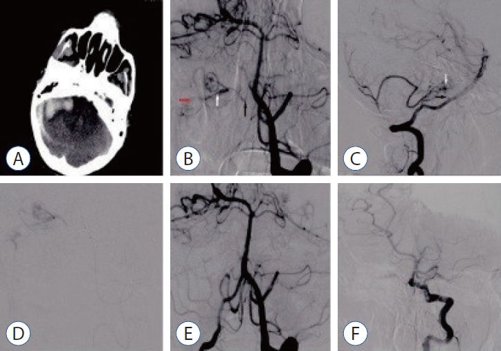
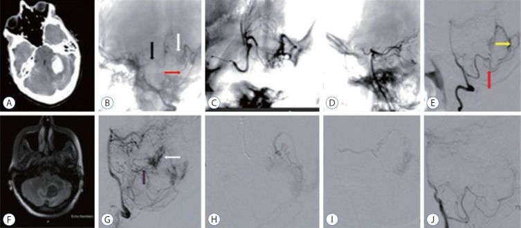
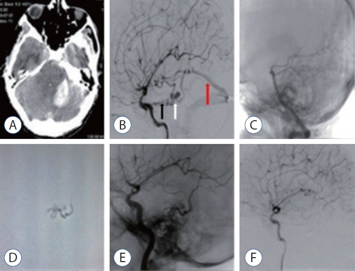
We evaluated the diagnosis, treatment, and long-term results of patients with dural arteriovenous fistula (dAVF), which is a very rare cause of posterior fossa hemorrhage.
This study included 15 patients who underwent endovascular, surgical, combined, or Gamma Knife treatments between 2012 and 2020. Demographics and clinical features, angiographic features, treatment modalities, and outcomes were analyzed.
The mean age of the patients was 40±17 years (range, 17-68), and 68% were men (11/15). Seven of the patients (46.6%) were in the age group of 50 years and older. While the mean Glasgow coma scale was 11.5±3.9 (range, 4-15), 46.3% presented with headache and 53.7% had stupor/coma. Four patients (26.6%) had only cerebellar hematoma and headache. All dAVFs had cortical venous drainage. In 11 patients (73.3%), the fistula was located in the tentorium and was the most common localization. Three patients (20%) had transverse and sigmoid sinus localizations, while one patient (6.7%) had dAVF located in the foramen magnum. Eighteen sessions were performed on the patients during endovascular treatment. Sixteen sessions (88.8%) were performed with the transarterial (TA) route, one session (5.5%) with the transvenous (TV) route, and one session (5.5%) with the TA+TV route. Surgery was performed in two patients (14.2%). One patient (7.1%) passed away. While there were nine patients (64.2%) with a Rankin score between 0 and 2, the total closure rate was 69.2% in the first year of control angiograms.
In the differential diagnosis of posterior fossa hemorrhages, the differential diagnosis of dAVFs, which is a very rare entity, should be considered, even in the middle and elderly age groups, in patients presenting with good clinical status and pure hematoma. The treatment of such patients can be done safely and effectively in a multidisciplinary manner with a good understanding of pathological vascular anatomy and appropriate endovascular treatment approaches.
我们评估了硬脑膜动静脉瘘(dAVF)患者的诊断、治疗及长期预后,dAVF是后颅窝出血的一种非常罕见的病因。
本研究纳入了2012年至2020年间接受血管内、手术、联合或伽玛刀治疗的15例患者。分析了人口统计学和临床特征、血管造影特征、治疗方式及预后。
患者的平均年龄为40±17岁(范围17 - 68岁),68%为男性(11/15)。7例患者(46.6%)年龄在50岁及以上。格拉斯哥昏迷量表平均分为11.5±3.9(范围4 - 15),46.3%的患者表现为头痛,53.7%的患者有昏迷/昏睡。4例患者(26.6%)仅有小脑血肿和头痛。所有dAVF均有皮质静脉引流。11例患者(73.3%)的瘘位于小脑幕,是最常见的部位。3例患者(20%)有横窦和乙状窦部位的病变,1例患者(6.7%)的dAVF位于枕骨大孔。血管内治疗期间对患者进行了18次操作。16次操作(88.8%)采用经动脉(TA)途径,1次操作(5.5%)采用经静脉(TV)途径,1次操作(5.5%)采用TA + TV途径。2例患者(14.2%)接受了手术。1例患者(7.1%)死亡。虽然9例患者(64.2%)的改良Rankin量表评分为0至2分,但在首次对照血管造影的第一年,总闭合率为69.2%。
在后颅窝出血的鉴别诊断中,即使在中老年人群中,对于临床表现良好且为单纯血肿的患者,也应考虑dAVF这种非常罕见病因的鉴别诊断。通过充分了解病理性血管解剖结构并采用适当的血管内治疗方法,多学科方式能够安全有效地治疗此类患者。