• 文献检索
  • 文档翻译
  • 深度研究
  • 学术资讯
  • Suppr Zotero 插件Zotero 插件
  • 邀请有礼
  • 套餐&价格
  • 历史记录
应用&插件
Suppr Zotero 插件Zotero 插件浏览器插件Mac 客户端Windows 客户端微信小程序
定价
高级版会员购买积分包购买API积分包
服务
文献检索文档翻译深度研究API 文档MCP 服务
关于我们
关于 Suppr公司介绍联系我们用户协议隐私条款
关注我们

Suppr 超能文献

核心技术专利:CN118964589B侵权必究
粤ICP备2023148730 号-1Suppr @ 2026

文献检索

告别复杂PubMed语法,用中文像聊天一样搜索,搜遍4000万医学文献。AI智能推荐,让科研检索更轻松。

立即免费搜索

文件翻译

保留排版,准确专业,支持PDF/Word/PPT等文件格式,支持 12+语言互译。

免费翻译文档

深度研究

AI帮你快速写综述,25分钟生成高质量综述,智能提取关键信息,辅助科研写作。

立即免费体验

在急性肺栓塞患者中,肺动脉直径与右心室功能障碍的超声心动图参数相关。

Pulmonary artery diameter correlates with echocardiographic parameters of right ventricular dysfunction in patients with acute pulmonary embolism.

作者信息

O'Corragain Oisin, Alashram Rami, Millio Gregory, Vanchiere Catherine, Hwang John Hojoon, Kumaran Maruti, Dass Chandra, Zhao Huaqing, Panero Joseph, Lakhter Vlad, Gupta Rohit, Bashir Riyaz, Cohen Gary, Jimenez David, Criner Gerard, Rali Parth

机构信息

Department of Thoracic Medicine and Surgery, Lewis Katz School of Medicine at Temple University, Philadelphia, PA, USA.

Department of Medicine, Lewis Katz School of Medicine at Temple University, Philadelphia, PA, USA.

出版信息

Lung India. 2023 Jul-Aug;40(4):306-311. doi: 10.4103/lungindia.lungindia_357_22.

DOI:10.4103/lungindia.lungindia_357_22
PMID:37417082
原文链接:https://pmc.ncbi.nlm.nih.gov/articles/PMC10401985/
Abstract

INTRODUCTION

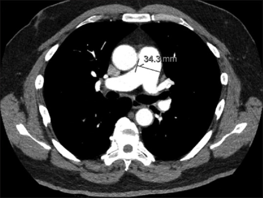
Right ventricular dysfunction (RVD) is a key component in the process of risk stratification in patients with acute pulmonary embolism (PE). Echocardiography remains the gold standard for RVD assessment, however, measures of RVD may be seen on CTPA imaging, including increased pulmonary artery diameter (PAD). The aim of our study was to evaluate the association between PAD and echocardiographic parameters of RVD in patients with acute PE.

METHODS

Retrospective analysis of patients diagnosed with acute PE was conducted at large academic center with an established pulmonary embolism response team (PERT). Patients with available clinical, imaging, and echocardiographic data were included. PAD was compared to echocardiographic markers of RVD. Statistical analysis was performed using the Student's t test, Chi-square test, or one-way analysis of variance (ANOVA); P < 0.05 was considered statistically significant.

RESULTS

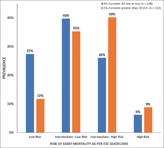
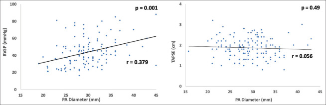
270 patients with acute PE were identified. Patients with a PAD >30 mm measured on CTPA had higher rates of RV dilation (73.1% vs 48.7%, P < 0.005), RV systolic dysfunction (65.4% vs 43.7%, P < 0.005), and RVSP >30 mmHg (90.2% vs 68%, P = 0.004), but not TAPSE ≤1.6 cm (39.1% vs 26.1%, P = 0.086). A weak increasing linear relationship between PAD and RVSP was noted (r = 0.379, P = 0.001).

CONCLUSIONS

Increased PAD in patients with acute PE was significantly associated with echocardiographic markers of RVD. Increased PAD on CTPA in acute PE can serve as a rapid prognostic tool and assist with PE risk stratification at the time of diagnosis, allowing rapid mobilization of a PERT team and appropriate resource utilization.

摘要

引言

右心室功能障碍(RVD)是急性肺栓塞(PE)患者风险分层过程中的关键组成部分。超声心动图仍是评估RVD的金标准,然而,在CTPA成像上也可见到RVD的表现,包括肺动脉直径(PAD)增加。我们研究的目的是评估急性PE患者中PAD与RVD超声心动图参数之间的关联。

方法

在一个设有肺栓塞应对团队(PERT)的大型学术中心,对诊断为急性PE的患者进行回顾性分析。纳入具有可用临床、影像学和超声心动图数据的患者。将PAD与RVD的超声心动图标志物进行比较。使用学生t检验、卡方检验或单因素方差分析(ANOVA)进行统计分析;P<0.05被认为具有统计学意义。

结果

共确定了270例急性PE患者。CTPA测量的PAD>30mm的患者右心室扩张率更高(73.1%对48.7%,P<0.005)、右心室收缩功能障碍发生率更高(65.4%对43.7%,P<0.005)以及右心室收缩压>30mmHg的比例更高(90.2%对68%,P=0.004),但三尖瓣环平面收缩期位移≤1.6cm的比例差异无统计学意义(39.1%对26.1%,P=0.086)。观察到PAD与右心室收缩压之间存在微弱的线性增加关系(r=0.379,P=0.001)。

结论

急性PE患者中PAD增加与RVD的超声心动图标志物显著相关。急性PE患者CTPA上PAD增加可作为一种快速的预后工具,并有助于诊断时的PE风险分层,从而能够迅速调动PERT团队并合理利用资源。

https://cdn.ncbi.nlm.nih.gov/pmc/blobs/be8c/10401985/5b6ea2f484cb/LI-40-306-g004.jpg
https://cdn.ncbi.nlm.nih.gov/pmc/blobs/be8c/10401985/692f4e99aaee/LI-40-306-g001.jpg
https://cdn.ncbi.nlm.nih.gov/pmc/blobs/be8c/10401985/97978a06e1c8/LI-40-306-g002.jpg
https://cdn.ncbi.nlm.nih.gov/pmc/blobs/be8c/10401985/0123796fb750/LI-40-306-g003.jpg
https://cdn.ncbi.nlm.nih.gov/pmc/blobs/be8c/10401985/5b6ea2f484cb/LI-40-306-g004.jpg

相似文献

1
Pulmonary artery diameter correlates with echocardiographic parameters of right ventricular dysfunction in patients with acute pulmonary embolism.在急性肺栓塞患者中,肺动脉直径与右心室功能障碍的超声心动图参数相关。
Lung India. 2023 Jul-Aug;40(4):306-311. doi: 10.4103/lungindia.lungindia_357_22.
2
Ventricular-arterial decoupling is associated with in-hospital adverse events in normotensive pulmonary embolism.室-动脉解耦与血压正常的肺栓塞住院期间不良事件相关。
Int J Cardiovasc Imaging. 2022 Dec;38(12):2655-2665. doi: 10.1007/s10554-022-02677-x. Epub 2022 Jul 15.
3
Predictive value of reduced pulmonary arterial elasticity in acute pulmonary embolism for right ventricular dysfunction.急性肺栓塞肺动脉弹性降低对右心功能障碍的预测价值。
J Thromb Thrombolysis. 2023 Nov;56(4):529-537. doi: 10.1007/s11239-023-02873-z. Epub 2023 Aug 7.
4
The risk factor age in normotensive patients with pulmonary embolism: Effectiveness of age in predicting submassive pulmonary embolism, cardiac injury, right ventricular dysfunction and elevated systolic pulmonary artery pressure in normotensive pulmonary embolism patients.血压正常的肺栓塞患者的危险因素年龄:年龄对预测血压正常的肺栓塞患者亚大面积肺栓塞、心脏损伤、右心室功能障碍及收缩期肺动脉压升高的有效性。
Exp Gerontol. 2015 Sep;69:116-21. doi: 10.1016/j.exger.2015.05.007. Epub 2015 May 15.
5
Echocardiographic Pattern of Acute Pulmonary Embolism: Analysis of 511 Consecutive Patients.急性肺栓塞的超声心动图表现:511例连续患者分析
J Am Soc Echocardiogr. 2016 Sep;29(9):907-13. doi: 10.1016/j.echo.2016.05.016. Epub 2016 Jul 15.
6
Prevalence of long-term right ventricular dysfunction after acute pulmonary embolism: a systematic review and meta-analysis.急性肺栓塞后长期右心室功能障碍的患病率:一项系统评价和荟萃分析。
EClinicalMedicine. 2023 Aug 11;62:102153. doi: 10.1016/j.eclinm.2023.102153. eCollection 2023 Aug.
7
Echocardiographic evaluation of pulmonary hypertension, right ventricular function, and right ventricular-pulmonary arterial coupling in patients with rheumatoid arthritis.超声心动图评价类风湿关节炎患者肺动脉高压、右心功能及右心-肺血管耦联。
Clin Rheumatol. 2021 Jul;40(7):2651-2656. doi: 10.1007/s10067-020-05544-z. Epub 2021 Jan 14.
8
Defining right ventricular dysfunction by echocardiography in normotensive patients with pulmonary embolism.超声心动图定义正常血压肺栓塞患者的右心功能障碍。
Pol Arch Intern Med. 2020 Sep 30;130(9):741-747. doi: 10.20452/pamw.15459. Epub 2020 Jun 24.
9
Right ventricular dysfunction in hemodynamically stable patients with acute pulmonary embolism.血流动力学稳定的急性肺栓塞患者的右心室功能障碍
Thromb Res. 2014 Apr;133(4):555-9. doi: 10.1016/j.thromres.2014.01.010. Epub 2014 Jan 14.
10
The value of CTPA for diagnosing acute pulmonary thromboembolism and the ensuing right ventricular dysfunction.CTPA在诊断急性肺血栓栓塞症及随之发生的右心室功能障碍方面的价值。
Cell Biochem Biophys. 2014 Jul;69(3):517-22. doi: 10.1007/s12013-014-9826-6.

本文引用的文献

1
Risk Stratification in Acute Pulmonary Embolism: The Latest Algorithms.急性肺栓塞的风险分层:最新算法。
Semin Respir Crit Care Med. 2021 Apr;42(2):183-198. doi: 10.1055/s-0041-1722898. Epub 2021 Feb 6.
2
Risk Stratification of Acute Pulmonary Embolism and Determining the Effect on Chronic Cardiopulmonary Complications: The REACH Study.急性肺栓塞的风险分层及对慢性心肺并发症影响的确定:REACH研究
TH Open. 2020 Mar 30;4(1):e45-e50. doi: 10.1055/s-0040-1708558. eCollection 2020 Jan.
3
Right ventricular dysfunction is associated with the development of chronic thromboembolic pulmonary hypertension but not with mortality post-acute pulmonary embolism.
右心室功能障碍与慢性血栓栓塞性肺动脉高压的发生有关,但与急性肺栓塞后的死亡率无关。
Medicine (Baltimore). 2019 Nov;98(48):e17953. doi: 10.1097/MD.0000000000017953.
4
2019 ESC Guidelines for the diagnosis and management of acute pulmonary embolism developed in collaboration with the European Respiratory Society (ERS).2019年欧洲心脏病学会(ESC)与欧洲呼吸学会(ERS)合作制定的急性肺栓塞诊断和管理指南。
Eur Heart J. 2020 Jan 21;41(4):543-603. doi: 10.1093/eurheartj/ehz405.
5
Septal bowing and pulmonary artery diameter on computed tomography pulmonary angiography are associated with short-term outcomes in patients with acute pulmonary embolism.计算机断层扫描肺动脉造影中的室间隔弯曲和肺动脉直径与急性肺栓塞患者的短期预后相关。
Emerg Radiol. 2019 Dec;26(6):623-630. doi: 10.1007/s10140-019-01709-9. Epub 2019 Aug 2.
6
Diagnostic Accuracy of Point-of-Care Ultrasound Performed by Pulmonary Critical Care Physicians for Right Ventricle Assessment in Patients With Acute Pulmonary Embolism.肺重症监护医师进行的床旁超声心动图对急性肺栓塞患者右心室评估的诊断准确性
Crit Care Med. 2017 Dec;45(12):2040-2045. doi: 10.1097/CCM.0000000000002723.
7
Estimation of right ventricular dysfunction by computed tomography pulmonary angiography: a valuable adjunct for evaluating the severity of acute pulmonary embolism.通过计算机断层扫描肺动脉造影评估右心室功能障碍:评估急性肺栓塞严重程度的一项有价值的辅助手段。
J Thromb Thrombolysis. 2017 Feb;43(2):271-278. doi: 10.1007/s11239-016-1438-0.
8
The efficacy of CT for detection of right ventricular dysfunction in acute pulmonary embolism, and comparison with cardiac biomarkers.CT检测急性肺栓塞时右心室功能障碍的效能及与心脏生物标志物的比较。
Jpn J Radiol. 2015 Aug;33(8):471-8. doi: 10.1007/s11604-015-0447-9. Epub 2015 Jun 29.
9
Predictive Value of Computed Tomography in Acute Pulmonary Embolism: Systematic Review and Meta-analysis.计算机断层扫描在急性肺栓塞中的预测价值:系统评价与荟萃分析
Am J Med. 2015 Jul;128(7):747-59.e2. doi: 10.1016/j.amjmed.2015.01.023. Epub 2015 Feb 11.
10
CT-base pulmonary artery measurement in the detection of pulmonary hypertension: a meta-analysis and systematic review.基于CT的肺动脉测量在肺动脉高压检测中的应用:一项荟萃分析与系统评价
Medicine (Baltimore). 2014 Dec;93(27):e256. doi: 10.1097/MD.0000000000000256.