• 文献检索
  • 文档翻译
  • 深度研究
  • 学术资讯
  • Suppr Zotero 插件Zotero 插件
  • 邀请有礼
  • 套餐&价格
  • 历史记录
应用&插件
Suppr Zotero 插件Zotero 插件浏览器插件Mac 客户端Windows 客户端微信小程序
定价
高级版会员购买积分包购买API积分包
服务
文献检索文档翻译深度研究API 文档MCP 服务
关于我们
关于 Suppr公司介绍联系我们用户协议隐私条款
关注我们

Suppr 超能文献

核心技术专利:CN118964589B侵权必究
粤ICP备2023148730 号-1Suppr @ 2026

文献检索

告别复杂PubMed语法,用中文像聊天一样搜索,搜遍4000万医学文献。AI智能推荐,让科研检索更轻松。

立即免费搜索

文件翻译

保留排版,准确专业,支持PDF/Word/PPT等文件格式,支持 12+语言互译。

免费翻译文档

深度研究

AI帮你快速写综述,25分钟生成高质量综述,智能提取关键信息,辅助科研写作。

立即免费体验

微创技术用于上颌窦外侧壁提升:小侧窗和一期手术——一项 2-5 年回顾性研究。

Minimally invasive techniques for lateral maxillary sinus floor elevation: small lateral window and one-stage surgery-a 2-5-year retrospective study.

机构信息

State Key Laboratory of Oral Diseases, West China Hospital of Stomatology, Sichuan University, Chengdu, China.

Department of Demography, Zhou Enlai School of Government, Nankai University, Tianjin, China.

出版信息

Int J Oral Sci. 2023 Jul 11;15(1):28. doi: 10.1038/s41368-023-00233-4.

DOI:10.1038/s41368-023-00233-4
PMID:37433766
原文链接:https://pmc.ncbi.nlm.nih.gov/articles/PMC10336061/
Abstract

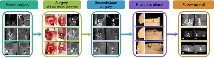
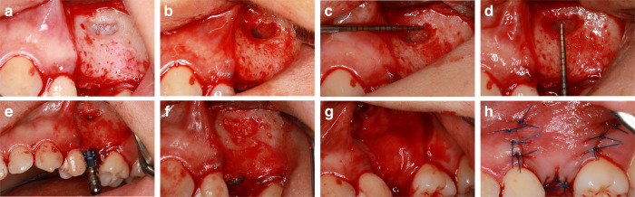
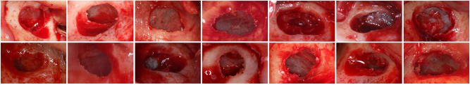
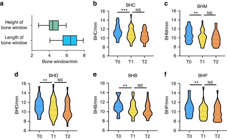
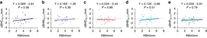
This study aimed to introduce a minimally invasive technique for maxillary sinus floor elevation using the lateral approach (lSFE) and to determine the factors that influence the stability of the grafted area in the sinus cavity. Thirty patients (30 implants) treated with lSFE using minimally invasive techniques from 2015 to 2019 were included in the study. Five aspects of the implant (central, mesial, distal, buccal, and palatal bone heights [BHs]) were measured using cone-beam computed tomography (CBCT) before implant surgery, immediately after surgery (T0), 6 months after surgery (T1), and at the last follow-up visit (T2). Patients' characteristics were collected. A small bone window (height, (4.40 ± 0.74) mm; length, (6.26 ± 1.03) mm) was prepared. No implant failed during the follow-up period (3.67 ± 1.75) years. Three of the 30 implants exhibited perforations. Changes in BH of the five aspects of implants showed strong correlations with each other and BH decreased dramatically before second-stage surgery. Residual bone height (RBH) did not significantly influence BH changes, whereas smoking status and type of bone graft materials were the potentially influential factors. During the approximate three-year observation period, lSFE with a minimally invasive technique demonstrated high implant survival rate and limited bone reduction in grafted area. In conclusion, lSFE using minimally invasive techniques was a viable treatment option. Patients who were nonsmokers and whose sinus cavity was filled with deproteinized bovine bone mineral (DBBM) had significantly limited bone resorption in grafted area.

摘要

本研究旨在介绍一种使用外侧入路(lSFE)的微创上颌窦底提升技术,并确定影响窦腔移植区稳定性的因素。2015 年至 2019 年期间,采用微创技术对 30 例患者(30 个种植体)进行 lSFE,将其纳入本研究。使用锥形束计算机断层扫描(CBCT)在种植术前、术后即刻(T0)、术后 6 个月(T1)和末次随访(T2)时分别测量种植体的五个方面(中央、近中、远中、颊侧和腭侧骨高度[BH])。收集患者特征。制备小骨窗(高度,(4.40±0.74)mm;长度,(6.26±1.03)mm)。在 3.67±1.75 年的随访期间,没有种植体失败。30 个种植体中有 3 个出现穿孔。五个方面的种植体 BH 变化之间具有很强的相关性,且在二期手术前 BH 显著下降。残留骨高度(RBH)对 BH 变化无显著影响,而吸烟状况和骨移植材料类型是潜在的影响因素。在大约 3 年的观察期内,微创技术的 lSFE 显示出较高的种植体存活率和有限的移植区骨质减少。结论:微创技术的 lSFE 是一种可行的治疗选择。非吸烟者和窦腔填充脱蛋白牛骨矿物质(DBBM)的患者,移植区骨质吸收明显受限。

https://cdn.ncbi.nlm.nih.gov/pmc/blobs/6452/10336061/832c006fa318/41368_2023_233_Fig6_HTML.jpg
https://cdn.ncbi.nlm.nih.gov/pmc/blobs/6452/10336061/162c37fd81f0/41368_2023_233_Fig1_HTML.jpg
https://cdn.ncbi.nlm.nih.gov/pmc/blobs/6452/10336061/8513a165e38c/41368_2023_233_Fig2_HTML.jpg
https://cdn.ncbi.nlm.nih.gov/pmc/blobs/6452/10336061/cfd174e6663e/41368_2023_233_Fig3_HTML.jpg
https://cdn.ncbi.nlm.nih.gov/pmc/blobs/6452/10336061/76e9c50e6a88/41368_2023_233_Fig4_HTML.jpg
https://cdn.ncbi.nlm.nih.gov/pmc/blobs/6452/10336061/80986cea4f21/41368_2023_233_Fig5_HTML.jpg
https://cdn.ncbi.nlm.nih.gov/pmc/blobs/6452/10336061/832c006fa318/41368_2023_233_Fig6_HTML.jpg

相似文献

1
Minimally invasive techniques for lateral maxillary sinus floor elevation: small lateral window and one-stage surgery-a 2-5-year retrospective study.微创技术用于上颌窦外侧壁提升:小侧窗和一期手术——一项 2-5 年回顾性研究。
Int J Oral Sci. 2023 Jul 11;15(1):28. doi: 10.1038/s41368-023-00233-4.
2
Influence of Lateral Windows with Decreased Vertical Height Following Maxillary Sinus Floor Augmentation: A 1-year Clinical and Radiographic Study.上颌窦底提升术后垂直高度降低的侧窗的影响:一项为期1年的临床和影像学研究。
Int J Oral Maxillofac Implants. 2018 May/Jun;33(3):661-670. doi: 10.11607/jomi.6213.
3
Changes in bone graft height and influencing factors after sinus floor augmentation by using the lateral window approach: A clinical retrospective study of 1 to 2 years.采用外侧开窗法行上颌窦底提升术后骨增量高度变化及其影响因素:一项1至2年的临床回顾性研究
J Prosthet Dent. 2023 Sep;130(3):362-368. doi: 10.1016/j.prosdent.2021.10.010. Epub 2021 Nov 29.
4
Radiographic outcomes of lateral sinus floor elevation with and without bone window repositioning: one-year results of a randomized controlled trial.伴或不伴骨窗复位的外侧上颌窦底提升的影像学结果:一项随机对照试验的一年结果
Int J Oral Maxillofac Surg. 2023 Feb;52(2):255-263. doi: 10.1016/j.ijom.2022.01.021. Epub 2022 Jul 1.
5
Radiographic outcomes of transcrestal and lateral sinus floor elevation: One-year results of a bi-center, parallel-arm randomized trial.经皮质和侧窦底提升的放射学结果:一项双中心、平行臂随机试验的一年结果。
Clin Oral Implants Res. 2019 Sep;30(9):910-919. doi: 10.1111/clr.13497. Epub 2019 Jul 10.
6
The comparative evaluation of transcrestal and lateral sinus floor elevation in sites with residual bone height ≤6 mm: A two-year prospective randomized study.经骨高度≤6mm 位点的经牙槽嵴顶和外侧窦底提升的对比评估:一项为期两年的前瞻性随机研究。
Clin Oral Implants Res. 2021 Feb;32(2):180-191. doi: 10.1111/clr.13688. Epub 2020 Dec 26.
7
Repositioning of the bone window in lateral sinus floor elevation with simultaneous implant placement: A retrospective radiographic study.同期种植术中应用骨窗侧位定位抬高颅后窝底:一项回顾性影像学研究。
Clin Oral Implants Res. 2022 Aug;33(8):816-833. doi: 10.1111/clr.13963. Epub 2022 Jun 25.
8
Long-term stability of autologous bone graft of intraoral origin after lateral sinus floor elevation with simultaneous implant placement.同期种植体植入术中经外侧窦底提升后取自口腔内的自体骨移植物的长期稳定性。
Clin Implant Dent Relat Res. 2018 Oct;20(5):713-721. doi: 10.1111/cid.12649. Epub 2018 Aug 24.
9
[Radiography study on osteotome sinus floor elevation with placed implant simultaneously with no graft augmentation].[不进行植骨增量同期植入种植体的骨凿上颌窦底提升术的影像学研究]
Beijing Da Xue Xue Bao Yi Xue Ban. 2020 Dec 24;53(1):95-101. doi: 10.19723/j.issn.1671-167X.2021.01.015.
10
Clinical and radiographic outcomes of reentry lateral sinus floor elevation after a complete membrane perforation.完全硬脑膜穿孔后再入侧窦底提升的临床和放射学结果。
Clin Implant Dent Relat Res. 2020 Oct;22(5):574-581. doi: 10.1111/cid.12932. Epub 2020 Sep 14.

引用本文的文献

1
Assessment of guided lateral maxillary sinus lift procedure with simultaneous implant placement using stereolithographic surgical guide: a randomized controlled clinical study.使用立体光刻手术导板评估引导式上颌窦外侧提升术同期种植体植入:一项随机对照临床研究。
Oral Maxillofac Surg. 2025 May 17;29(1):102. doi: 10.1007/s10006-025-01399-3.
2
Biomechanical analysis of maxillary posterior three unit bridge supported misial straight implant and distal tilted implant.上颌后牙区近中直形种植体和远中倾斜种植体支持的三单位桥的生物力学分析
Front Bioeng Biotechnol. 2025 Feb 25;13:1546656. doi: 10.3389/fbioe.2025.1546656. eCollection 2025.

本文引用的文献

1
Does new bone formation vary in different sites within the same maxillary sinus after lateral augmentation? A prospective histomorphometric study.上颌窦侧壁增宽术后同一窦内不同部位新骨形成是否存在差异?一项前瞻性组织形态计量学研究。
Clin Oral Implants Res. 2022 Mar;33(3):322-332. doi: 10.1111/clr.13891. Epub 2022 Jan 12.
2
Changes in bone graft height and influencing factors after sinus floor augmentation by using the lateral window approach: A clinical retrospective study of 1 to 2 years.采用外侧开窗法行上颌窦底提升术后骨增量高度变化及其影响因素:一项1至2年的临床回顾性研究
J Prosthet Dent. 2023 Sep;130(3):362-368. doi: 10.1016/j.prosdent.2021.10.010. Epub 2021 Nov 29.
3
Risk factors for sinus membrane perforation during lateral window maxillary sinus floor elevation surgery: A retrospective study.
鼻窦侧壁开窗提升上颌窦底术中发生窦膜穿孔的危险因素:一项回顾性研究。
Clin Implant Dent Relat Res. 2021 Dec;23(6):812-820. doi: 10.1111/cid.13052. Epub 2021 Nov 8.
4
Individualized plasticity autograft mimic with efficient bioactivity inducing osteogenesis.具有高效生物活性诱导成骨作用的个体化塑性自体移植物模拟物。
Int J Oral Sci. 2021 Apr 12;13(1):14. doi: 10.1038/s41368-021-00120-w.
5
The assessment of the effect of the size of lateral-antrostomy in graftless balloon elevation of the maxillary sinus membrane with simultaneous implant placement (a randomized controlled clinical trial).评价同期种植体植入时无移植物球囊上颌窦提升中侧窦造口大小对窦膜提升效果的影响(一项随机对照临床试验)。
Clin Implant Dent Relat Res. 2021 Feb;23(1):31-42. doi: 10.1111/cid.12983. Epub 2021 Feb 17.
6
Early bone formation in mini-lateral window sinus floor elevation with simultaneous implant placement: An in vivo experimental study.同期种植微型外侧提升窦底骨增量的早期成骨:一项体内实验研究。
Clin Oral Implants Res. 2021 Apr;32(4):448-459. doi: 10.1111/clr.13714. Epub 2021 Feb 9.
7
Titanium mesh for bone augmentation in oral implantology: current application and progress.钛网在口腔种植学中的骨增量应用:现状与进展。
Int J Oral Sci. 2020 Dec 30;12(1):37. doi: 10.1038/s41368-020-00107-z.
8
Minimally Invasive Management of Implant-Supported Rehabilitation in the Posterior Maxilla, Part II. Surgical Techniques and Decision Tree.上颌后牙区种植支持修复的微创治疗,第二部分。手术技术与决策树
Int J Periodontics Restorative Dent. 2020 May/Jun;40(3):e95-e102. doi: 10.11607/prd.4498.
9
Quantitative Assessment of the Edentulous Posterior Maxilla for Implant Therapy: A Retrospective Cone Beam Computed Tomographic Study.牙列缺失后上颌骨种植治疗的定量评估:一项回顾性锥形束计算机断层扫描研究
J Maxillofac Oral Surg. 2020 Mar;19(1):125-130. doi: 10.1007/s12663-019-01236-7. Epub 2019 May 9.
10
A Less-Invasive Window Design for Lateral Wall Maxillary Sinus Augmentations.一种用于上颌窦外侧壁增高术的微创开窗设计
Int J Periodontics Restorative Dent. 2019 Nov/Dec;39(6):855-861. doi: 10.11607/prd.4206.