• 文献检索
  • 文档翻译
  • 深度研究
  • 学术资讯
  • Suppr Zotero 插件Zotero 插件
  • 邀请有礼
  • 套餐&价格
  • 历史记录
应用&插件
Suppr Zotero 插件Zotero 插件浏览器插件Mac 客户端Windows 客户端微信小程序
定价
高级版会员购买积分包购买API积分包
服务
文献检索文档翻译深度研究API 文档MCP 服务
关于我们
关于 Suppr公司介绍联系我们用户协议隐私条款
关注我们

Suppr 超能文献

核心技术专利:CN118964589B侵权必究
粤ICP备2023148730 号-1Suppr @ 2026

文献检索

告别复杂PubMed语法,用中文像聊天一样搜索,搜遍4000万医学文献。AI智能推荐,让科研检索更轻松。

立即免费搜索

文件翻译

保留排版,准确专业,支持PDF/Word/PPT等文件格式,支持 12+语言互译。

免费翻译文档

深度研究

AI帮你快速写综述,25分钟生成高质量综述,智能提取关键信息,辅助科研写作。

立即免费体验

上颌窦侧壁增宽术后同一窦内不同部位新骨形成是否存在差异?一项前瞻性组织形态计量学研究。

Does new bone formation vary in different sites within the same maxillary sinus after lateral augmentation? A prospective histomorphometric study.

机构信息

Department of Medical, Surgical and Health Sciences, University of Trieste, Trieste, Italy.

Department of Health Sciences, University 'Magna Graecia', Catanzaro, Italy.

出版信息

Clin Oral Implants Res. 2022 Mar;33(3):322-332. doi: 10.1111/clr.13891. Epub 2022 Jan 12.

DOI:10.1111/clr.13891
PMID:34978096
原文链接:https://pmc.ncbi.nlm.nih.gov/articles/PMC9306466/
Abstract

OBJECTIVE

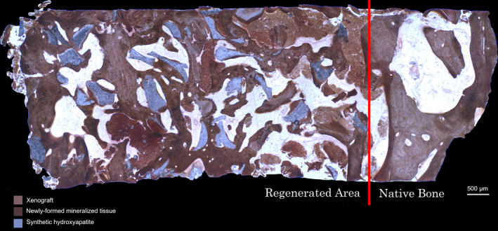
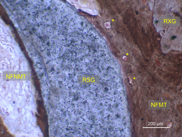
The aim of this study was to evaluate histomorphometric outcomes of lateral maxillary sinus augmentation in different areas of the same cavity and to correlate results to bucco-palatal sinus width (SW) and residual bone height (RBH).

MATERIAL AND METHODS

Patients needing maxillary sinus floor elevation (RBH <5 mm) to insert two nonadjacent implants were treated with lateral augmentation using a composite graft. Six months later, two bone-core biopsies (mesial/distal) were retrieved in implant insertion sites. SW and RBH were measured on cone beam computed tomography, and correlations between histomorphometric and anatomical parameters were evaluated by multivariate linear regression analysis.

RESULTS

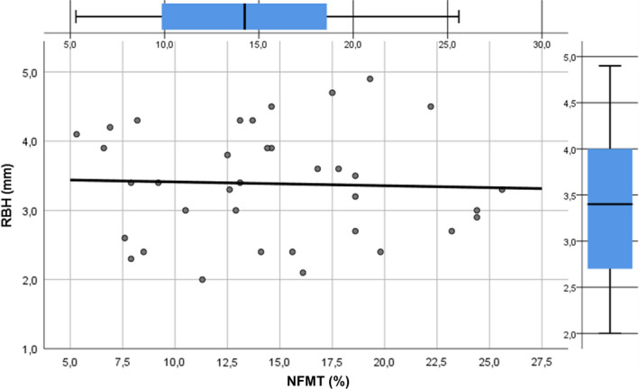
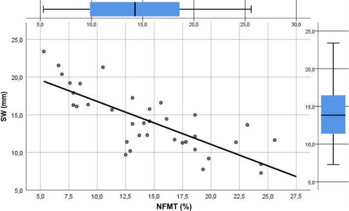
Twenty patients underwent sinus augmentation, and eighteen were included in the final analysis (two dropouts for membrane perforation). Mean newly formed mineralized tissue percentage (%NFMT) after 6 months in mesial and distal sites was 17.5 ± 4.7 and 11.6 ± 4.7, respectively (p = .0004). Multivariate linear regression showed a strong negative correlation between SW and %NFMT (β coefficient=-.774, p < .0001) and no correlation between RBH and %NFMT (β coefficient =-.038, p = .825).

CONCLUSIONS

The present study confirms that %NFMT after lateral sinus augmentation occurs at different rates in different anatomical areas of the same maxillary sinus, showing a strong negative correlation with SW, whereas no influence of RBH was observed. Clinicians should regard SW as a guide for graft selection and to decide duration of the healing period. Researchers should consider SW as a predictor variable, when comparing regenerative outcomes of different biomaterials by using maxillary sinus as an experimental model.

摘要

目的

本研究旨在评估同一上颌窦不同区域的外侧上颌窦提升术的组织形态计量学结果,并将结果与颊-腭向窦宽(SW)和剩余骨高度(RBH)相关联。

材料和方法

需要进行上颌窦底提升(RBH <5 mm)以植入两个不相邻种植体的患者,采用复合移植物进行外侧增强。6 个月后,在植入部位取出两个骨芯活检(近中/远中)。在锥形束计算机断层扫描上测量 SW 和 RBH,并通过多元线性回归分析评估组织形态计量学和解剖参数之间的相关性。

结果

20 名患者接受了鼻窦增强,其中 18 名患者进入了最终分析(两名因膜穿孔脱落)。近中部位和远中部位 6 个月后新形成的矿化组织百分比(%NFMT)分别为 17.5 ± 4.7 和 11.6 ± 4.7(p = 0.0004)。多元线性回归显示,SW 与 %NFMT 之间存在强烈的负相关(β系数=-.774,p < 0.0001),而 RBH 与 %NFMT 之间无相关性(β系数=-.038,p = 0.825)。

结论

本研究证实,外侧鼻窦提升后,同一上颌窦不同解剖区域的 %NFMT 以不同的速率发生,与 SW 呈强烈的负相关,而 RBH 无影响。临床医生应将 SW 作为指导,选择移植物并决定愈合期的持续时间。研究人员在使用上颌窦作为实验模型比较不同生物材料的再生结果时,应将 SW 视为预测变量。

https://cdn.ncbi.nlm.nih.gov/pmc/blobs/e967/9306466/9e5e6ad23500/CLR-33-322-g004.jpg
https://cdn.ncbi.nlm.nih.gov/pmc/blobs/e967/9306466/cf6c9269b67e/CLR-33-322-g002.jpg
https://cdn.ncbi.nlm.nih.gov/pmc/blobs/e967/9306466/4490485fd024/CLR-33-322-g001.jpg
https://cdn.ncbi.nlm.nih.gov/pmc/blobs/e967/9306466/37a10b8691c2/CLR-33-322-g003.jpg
https://cdn.ncbi.nlm.nih.gov/pmc/blobs/e967/9306466/9e5e6ad23500/CLR-33-322-g004.jpg

相似文献

1
Does new bone formation vary in different sites within the same maxillary sinus after lateral augmentation? A prospective histomorphometric study.上颌窦侧壁增宽术后同一窦内不同部位新骨形成是否存在差异?一项前瞻性组织形态计量学研究。
Clin Oral Implants Res. 2022 Mar;33(3):322-332. doi: 10.1111/clr.13891. Epub 2022 Jan 12.
2
New bone formation after transcrestal sinus floor elevation was influenced by sinus cavity dimensions: A prospective histologic and histomorphometric study.经牙槽嵴窦底提升后新骨形成受窦腔尺寸影响:一项前瞻性组织学和组织形态计量学研究。
Clin Oral Implants Res. 2018 May;29(5):465-479. doi: 10.1111/clr.13144. Epub 2018 Mar 23.
3
Influence of Maxillary Sinus Width on New Bone Formation After Transcrestal Sinus Floor Elevation: A Proof-of-Concept Prospective Cohort Study.经牙槽嵴上颌窦底提升术后上颌窦宽度对新骨形成的影响:一项概念验证性前瞻性队列研究。
Implant Dent. 2017 Apr;26(2):209-216. doi: 10.1097/ID.0000000000000554.
4
The effect of anatomy on osteogenesis after maxillary sinus floor augmentation: a radiographic and histological analysis.上颌窦底提升后解剖结构对成骨的影响:影像学和组织学分析。
Clin Oral Investig. 2021 Sep;25(9):5197-5204. doi: 10.1007/s00784-021-03827-6. Epub 2021 Feb 11.
5
[Radiography study on osteotome sinus floor elevation with placed implant simultaneously with no graft augmentation].[不进行植骨增量同期植入种植体的骨凿上颌窦底提升术的影像学研究]
Beijing Da Xue Xue Bao Yi Xue Ban. 2020 Dec 24;53(1):95-101. doi: 10.19723/j.issn.1671-167X.2021.01.015.
6
Sinus floor augmentation using crestal approach in conjunction with hydroxyapatite/cross-linked collagen sponge: A pilot study.经牙槽嵴顶入路并用羟磷灰石/交联胶原海绵行牙槽嵴增高术:一项初步研究。
Clin Implant Dent Relat Res. 2023 Oct;25(5):974-983. doi: 10.1111/cid.13236. Epub 2023 Jun 8.
7
Using Platelet-Rich Fibrin in Combination With Allograft Bone Particles Can Induce Bone Formation in Maxillary Sinus Augmentation.富血小板纤维蛋白与同种异体骨颗粒联合使用可促进上颌窦提升中的骨形成。
J Oral Maxillofac Surg. 2023 Jul;81(7):904-912. doi: 10.1016/j.joms.2023.03.015. Epub 2023 Mar 30.
8
Effect of Maxillary Sinus Anatomy on Bone Gain After Lateral Window Sinus Floor Elevation: A Case-Control Study.上颌窦解剖结构对侧壁开窗式上颌窦底提升后骨增量的影响:一项病例对照研究。
Int J Oral Maxillofac Implants. 2023 Mar-Apr;38(2):338-346. doi: 10.11607/jomi.9980.
9
Comprehensive sinus contour classification and its characteristics from radiographic examination: a cross-sectional study.基于影像学检查的鼻窦轮廓综合分类及其特征:一项横断面研究。
BMC Oral Health. 2024 Aug 30;24(1):1021. doi: 10.1186/s12903-024-04707-7.
10
The influence of the bucco-palatal distance on sinus augmentation outcomes.颊腭距离对鼻窦提升效果的影响。
J Periodontol. 2010 Jul;81(7):1041-50. doi: 10.1902/jop.2010.090686.

引用本文的文献

1
New bone formation of biphasic calcium phosphate bone substitute material: a systematic review and network meta-analysis of randomized controlled trials (RCTs).双相磷酸钙骨替代材料的新骨形成:随机对照试验(RCTs)的系统评价和网状Meta分析
Int J Implant Dent. 2025 Jul 15;11(1):47. doi: 10.1186/s40729-025-00636-4.
2
Finite Element Analysis of Functionally Loaded Subperiosteal Implants Evaluated on a Realistic Model Reproducing Severe Atrophic Jaws.在重现严重萎缩性颌骨的真实模型上评估功能负载骨膜下种植体的有限元分析
Methods Protoc. 2025 Jan 18;8(1):8. doi: 10.3390/mps8010008.
3
Biomechanical Basis for Bone Healing and Osseointegration of Implants in Sinus Grafts.

本文引用的文献

1
Radiographic and Histomorphometric Evaluation of Biomaterials Used for Lateral Sinus Augmentation: A Systematic Review on the Effect of Residual Bone Height and Vertical Graft Size on New Bone Formation and Graft Shrinkage.用于外侧窦增高的生物材料的影像学和组织形态计量学评估:关于剩余骨高度和垂直植骨尺寸对新骨形成和植骨收缩影响的系统评价
J Clin Med. 2021 Oct 27;10(21):4996. doi: 10.3390/jcm10214996.
2
The effect of anatomy on osteogenesis after maxillary sinus floor augmentation: a radiographic and histological analysis.上颌窦底提升后解剖结构对成骨的影响:影像学和组织学分析。
Clin Oral Investig. 2021 Sep;25(9):5197-5204. doi: 10.1007/s00784-021-03827-6. Epub 2021 Feb 11.
3
窦提升植骨术中骨愈合及种植体骨结合的生物力学基础
Clin Implant Dent Relat Res. 2025 Feb;27(1):e13424. doi: 10.1111/cid.13424. Epub 2024 Dec 5.
4
Minimally invasive techniques for lateral maxillary sinus floor elevation: small lateral window and one-stage surgery-a 2-5-year retrospective study.微创技术用于上颌窦外侧壁提升:小侧窗和一期手术——一项 2-5 年回顾性研究。
Int J Oral Sci. 2023 Jul 11;15(1):28. doi: 10.1038/s41368-023-00233-4.
5
Residual Bone Height and New Bone Formation after Maxillary Sinus Augmentation Procedure Using Biomaterials: A Network Meta-Analysis of Clinical Trials.使用生物材料进行上颌窦提升术后的剩余骨高度和新骨形成:一项临床试验的网状Meta分析
Materials (Basel). 2023 Feb 6;16(4):1376. doi: 10.3390/ma16041376.
6
Combined Application of Dentin Noncollagenous Proteins and Odontogenic Biphasic Calcium Phosphate in Rabbit Maxillary Sinus Lifting.牙本质非胶原蛋白与牙源性双相磷酸钙在兔上颌窦提升中的联合应用。
Tissue Eng Regen Med. 2023 Feb;20(1):93-109. doi: 10.1007/s13770-022-00502-z. Epub 2022 Dec 24.
7
Healing Patterns of Non-Collagenated Bovine and Collagenated Porcine Xenografts Used for Sinus Floor Elevation: A Histological Study in Rabbits.用于上颌窦底提升的非胶原化牛异种移植物和胶原化猪异种移植物的愈合模式:一项在兔身上的组织学研究
J Funct Biomater. 2022 Dec 5;13(4):276. doi: 10.3390/jfb13040276.
8
Bone Graft Packing and Its Association with Bone Regeneration in Maxillary Sinus Floor Augmentations: Histomorphometric Analysis of Human Biopsies.骨移植填充及其在上颌窦底提升术中与骨再生的关系:人体活检组织形态计量学分析
Biology (Basel). 2022 Sep 30;11(10):1431. doi: 10.3390/biology11101431.
9
Clinical and radiographic outcomes following transcrestal maxillary sinus floor elevation with injectable xenogenous bone substitute in gel form: a prospective multicenter study.经皮上颌窦底提升同期注射型凝胶状异种骨移植的临床及影像学效果:一项前瞻性多中心研究。
Int J Implant Dent. 2022 Jul 22;8(1):32. doi: 10.1186/s40729-022-00431-5.
10
Intraoperative complications and early implant failure after transcrestal sinus floor elevation with residual bone height ≤5 mm: A retrospective multicenter study.经窦提升术治疗剩余骨高度 ≤5 mm 时的术中并发症和早期种植体失败:一项回顾性多中心研究。
Clin Oral Implants Res. 2022 Aug;33(8):783-791. doi: 10.1111/clr.13959. Epub 2022 May 29.
Relative performance of various biomaterials used for maxillary sinus augmentation: A Bayesian network meta-analysis.
各种用于上颌窦提升的生物材料的相对性能:贝叶斯网状荟萃分析。
Clin Oral Implants Res. 2021 Feb;32(2):135-153. doi: 10.1111/clr.13690. Epub 2021 Jan 6.
4
The Influence of Residual Alveolar Bone Height on Graft Composition after Maxillary Sinus Augmentation Using Two Different Xenografts: A Histomorphometric Comparative Study.两种不同异种骨在上颌窦提升后残余牙槽骨高度对植骨成分的影响:一项组织形态计量学比较研究
Materials (Basel). 2020 Nov 11;13(22):5093. doi: 10.3390/ma13225093.
5
Osteogenic capacity of the sinus membrane following maxillary sinus augmentation procedures: A systematic review.上颌窦提升术后窦膜成骨能力:系统评价。
Int J Oral Implantol (Berl). 2020;13(3):213-232.
6
The vertical course of bone regeneration in maxillary sinus floor augmentations: A histomorphometric analysis of human biopsies.上颌窦底骨增量的垂直骨再生过程:对人类活检样本的组织形态计量学分析。
J Periodontol. 2021 Feb;92(2):263-272. doi: 10.1002/JPER.19-0656. Epub 2020 Aug 19.
7
Minimally Invasive Management of Implant-Supported Rehabilitation in the Posterior Maxilla, Part II. Surgical Techniques and Decision Tree.上颌后牙区种植支持修复的微创治疗,第二部分。手术技术与决策树
Int J Periodontics Restorative Dent. 2020 May/Jun;40(3):e95-e102. doi: 10.11607/prd.4498.
8
Algae-derived hydroxyapatite behavior as bone biomaterial in comparison with anorganic bovine bone: A split-mouth clinical, radiological, and histologic randomized study in humans.藻酸盐衍生羟磷灰石作为骨生物材料的行为与无机牛骨的比较:一项在人类中进行的随机分组、临床、放射学和组织学的口腔内研究。
Clin Oral Implants Res. 2020 Jun;31(6):536-548. doi: 10.1111/clr.13590. Epub 2020 Mar 6.
9
PRGF as adjunct to DBB in maxillary sinus floor augmentation: histological results of a pilot split-mouth study.富血小板纤维蛋白作为上颌窦底提升术中双皮质骨块移植的辅助材料:一项初步的双侧对照研究的组织学结果
Int J Implant Dent. 2019 Apr 1;5(1):14. doi: 10.1186/s40729-019-0166-6.
10
Sinus floor elevation using particulate PLGA-coated biphasic calcium phosphate bone graft substitutes: A prospective histological and radiological study.使用颗粒状 PLGA 涂层双相磷酸钙骨移植物替代物进行鼻窦底提升:一项前瞻性组织学和影像学研究。
Clin Implant Dent Relat Res. 2019 Oct;21(5):895-902. doi: 10.1111/cid.12741. Epub 2019 Mar 20.