• 文献检索
  • 文档翻译
  • 深度研究
  • 学术资讯
  • Suppr Zotero 插件Zotero 插件
  • 邀请有礼
  • 套餐&价格
  • 历史记录
应用&插件
Suppr Zotero 插件Zotero 插件浏览器插件Mac 客户端Windows 客户端微信小程序
定价
高级版会员购买积分包购买API积分包
服务
文献检索文档翻译深度研究API 文档MCP 服务
关于我们
关于 Suppr公司介绍联系我们用户协议隐私条款
关注我们

Suppr 超能文献

核心技术专利:CN118964589B侵权必究
粤ICP备2023148730 号-1Suppr @ 2026

文献检索

告别复杂PubMed语法,用中文像聊天一样搜索,搜遍4000万医学文献。AI智能推荐,让科研检索更轻松。

立即免费搜索

文件翻译

保留排版,准确专业,支持PDF/Word/PPT等文件格式,支持 12+语言互译。

免费翻译文档

深度研究

AI帮你快速写综述,25分钟生成高质量综述,智能提取关键信息,辅助科研写作。

立即免费体验

基于 Elekta MRI-直线加速器联盟图像分析工作组的建议,以提高 ADC 推导的可重复性。

Recommendations for improved reproducibility of ADC derivation on behalf of the Elekta MRI-linac consortium image analysis working group.

机构信息

Laboratory of Radiation Physics, Department of Oncology, Odense University Hospital, Kløvervænget 19, 5000 Odense, Denmark; Department of Clinical Research, University of Southern Denmark, J.B. Winsløws Vej 19.3, 5000 Odense Denmark.

Department of Radiation Oncology, Radboud University Medical Centre, P.O. Box 9101, 6500 HB Nijmegen, The Netherlands.

出版信息

Radiother Oncol. 2023 Sep;186:109803. doi: 10.1016/j.radonc.2023.109803. Epub 2023 Jul 10.

DOI:10.1016/j.radonc.2023.109803
PMID:37437609
原文链接:https://pmc.ncbi.nlm.nih.gov/articles/PMC11197850/
Abstract

BACKGROUND AND PURPOSE

The apparent diffusion coefficient (ADC), a potential imaging biomarker for radiotherapy response, needs to be reproducible before translation into clinical use. The aim of this study was to evaluate the multi-centre delineation- and calculation-related ADC variation and give recommendations to minimize it.

MATERIALS AND METHODS

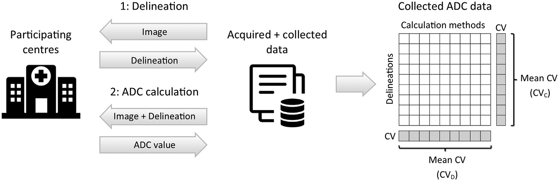
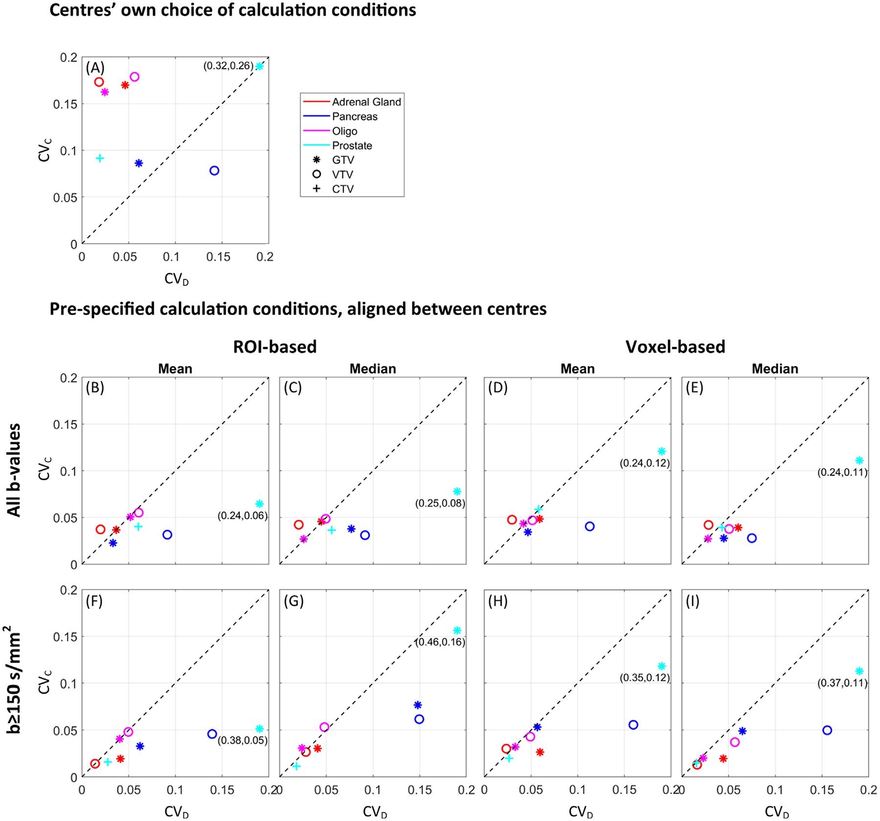
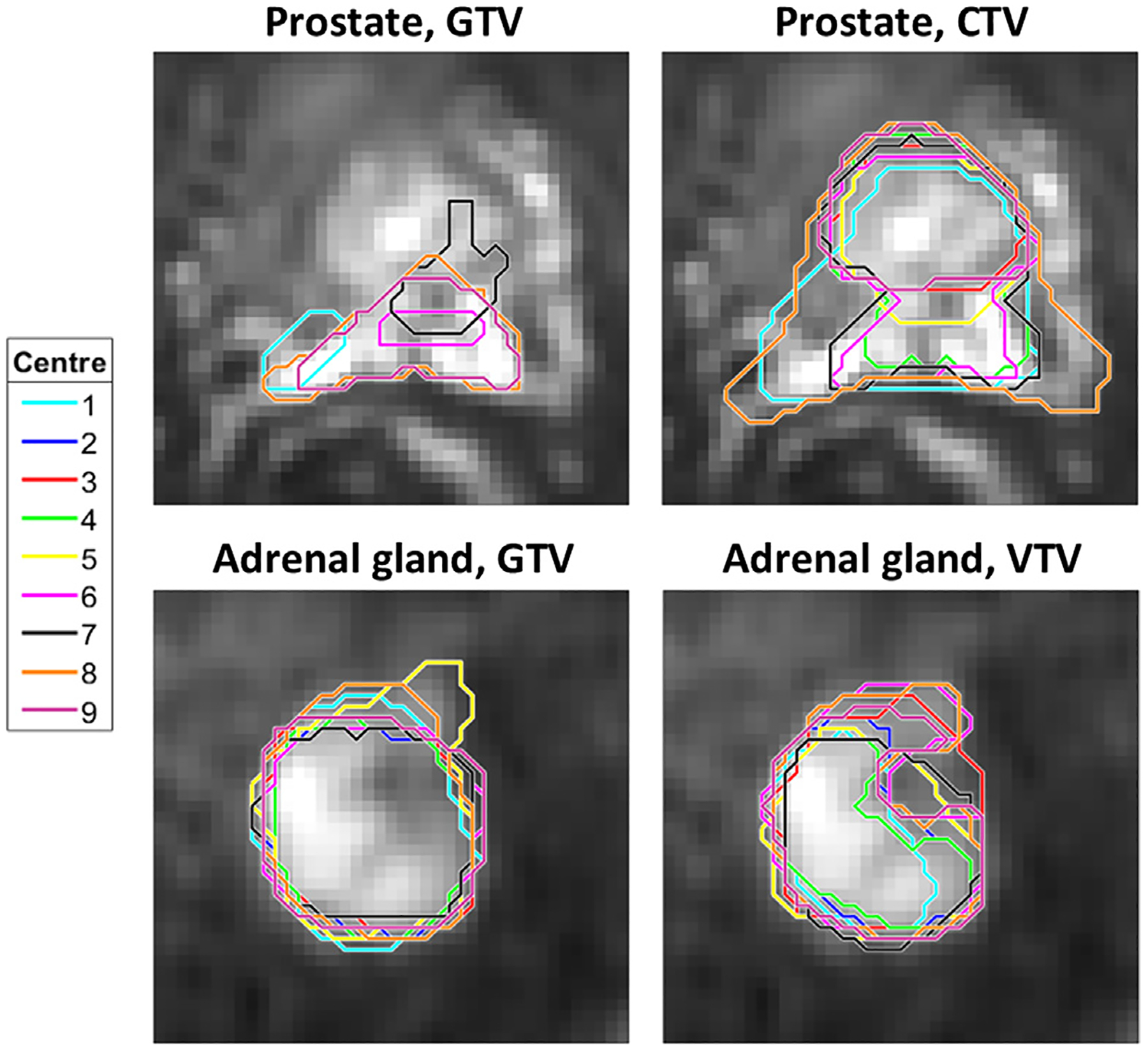
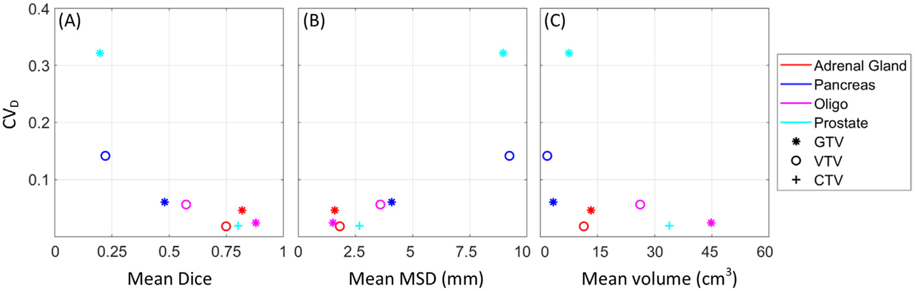
Nine centres received identical diffusion-weighted and anatomical magnetic resonance images of different cancerous tumours (adrenal gland, pelvic oligo metastasis, pancreas, and prostate). All centres delineated the gross tumour volume (GTV), clinical target volume (CTV), and viable tumour volume (VTV), and calculated ADCs using both their local calculation methods and each of the following calculation conditions: b-values 0-500 vs. 150-500 s/mm, region-of-interest (ROI)-based vs. voxel-based calculation, and mean vs. median. ADC variation was assessed using the mean coefficient of variation across delineations (CV) and calculation methods (CV). Absolute ADC differences between calculation conditions were evaluated using Friedman's test. Recommendations for ADC calculation were formulated based on observations and discussions within the Elekta MRI-linac consortium image analysis working group.

RESULTS

The median (range) CV and CV were 0.06 (0.02-0.32) and 0.17 (0.08-0.26), respectively. The ADC estimates differed 18% between b-value sets and 4% between ROI/voxel-based calculation (p-values < 0.01). No significant difference was observed between mean and median (p = 0.64). Aligning calculation conditions between centres reduced CV to 0.04 (0.01-0.16). CV was comparable between ROI types.

CONCLUSION

Overall, calculation methods had a larger impact on ADC reproducibility compared to delineation. Based on the results, significant sources of variation were identified, which should be considered when initiating new studies, in particular multi-centre investigations.

摘要

背景与目的

表观扩散系数(ADC)是一种潜在的放疗反应的影像学生物标志物,在转化为临床应用之前需要具有可重复性。本研究的目的是评估多中心勾画和计算相关 ADC 变化,并提出建议以最小化这种变化。

材料与方法

九个中心接收了不同癌症肿瘤(肾上腺、盆腔寡转移、胰腺和前列腺)的相同弥散加权和解剖磁共振图像。所有中心均勾画了大体肿瘤体积(GTV)、临床靶区体积(CTV)和存活肿瘤体积(VTV),并使用各自的局部计算方法和以下计算条件计算 ADC:b 值 0-500 与 150-500 s/mm、基于感兴趣区(ROI)与基于体素的计算、均值与中位数。通过勾画和计算方法的跨勾画变异系数(CV)和均值 CV 评估 ADC 变化。使用 Friedman 检验评估不同计算条件下的绝对 ADC 差异。根据 Elekta MRI-直线加速器联盟图像分析工作组的观察和讨论,制定了 ADC 计算建议。

结果

中位(范围)CV 和 CV 分别为 0.06(0.02-0.32)和 0.17(0.08-0.26)。b 值集之间的 ADC 估计值差异为 18%,ROI/体素计算之间的差异为 4%(p 值均小于 0.01)。均值和中位数之间无显著差异(p=0.64)。中心之间的计算条件对齐将 CV 降低至 0.04(0.01-0.16)。ROI 类型之间的 CV 相似。

结论

总体而言,与勾画相比,计算方法对 ADC 重现性的影响更大。基于这些结果,确定了显著的变异来源,在启动新研究时,特别是在多中心研究时,应考虑这些来源。

https://cdn.ncbi.nlm.nih.gov/pmc/blobs/ff37/11197850/9511b7eaef79/nihms-1996513-f0004.jpg
https://cdn.ncbi.nlm.nih.gov/pmc/blobs/ff37/11197850/4fff3b262f9d/nihms-1996513-f0001.jpg
https://cdn.ncbi.nlm.nih.gov/pmc/blobs/ff37/11197850/354d2c5d3e42/nihms-1996513-f0002.jpg
https://cdn.ncbi.nlm.nih.gov/pmc/blobs/ff37/11197850/8a0138876fd9/nihms-1996513-f0003.jpg
https://cdn.ncbi.nlm.nih.gov/pmc/blobs/ff37/11197850/9511b7eaef79/nihms-1996513-f0004.jpg

相似文献

1
Recommendations for improved reproducibility of ADC derivation on behalf of the Elekta MRI-linac consortium image analysis working group.基于 Elekta MRI-直线加速器联盟图像分析工作组的建议,以提高 ADC 推导的可重复性。
Radiother Oncol. 2023 Sep;186:109803. doi: 10.1016/j.radonc.2023.109803. Epub 2023 Jul 10.
2
Prostate diffusion-weighted imaging (DWI) in MR-guided radiotherapy: Reproducibility assessment on 1.5 T MR-Linac and 1.5 T MR-simulator.MR 引导放疗中的前列腺弥散加权成像(DWI):1.5T MR-Linac 和 1.5T MR 模拟器上的可重复性评估。
Magn Reson Imaging. 2024 Sep;111:47-56. doi: 10.1016/j.mri.2024.03.020. Epub 2024 Mar 19.
3
Longitudinal assessment of diffusion-weighted imaging during magnetic resonance-guided radiotherapy in head and neck cancer.头颈部癌磁共振引导放疗期间扩散加权成像的纵向评估
Radiat Oncol. 2025 Jan 29;20(1):15. doi: 10.1186/s13014-025-02589-9.
4
Apparent diffusion coefficients in GEC ESTRO target volumes for image guided adaptive brachytherapy of locally advanced cervical cancer.局部晚期宫颈癌图像引导自适应近距离治疗中 GEC ESTRO 靶区的表观扩散系数。
Acta Oncol. 2010 Oct;49(7):978-83. doi: 10.3109/0284186X.2010.500619.
5
Impact of measurement parameters on apparent diffusion coefficient quantification in diffusion-weighted-magnetic resonance imaging.测量参数对磁共振扩散加权成像中表观扩散系数定量的影响。
Invest Radiol. 2015 Jan;50(1):46-56. doi: 10.1097/RLI.0000000000000095.
6
Assessment and quantification of sources of variability in breast apparent diffusion coefficient (ADC) measurements at diffusion weighted imaging.扩散加权成像中乳腺表观扩散系数(ADC)测量变异性来源的评估与量化
Eur J Radiol. 2015 Sep;84(9):1729-36. doi: 10.1016/j.ejrad.2015.05.032. Epub 2015 Jun 1.
7
Impact of post-processing methods on apparent diffusion coefficient values.后处理方法对表观扩散系数值的影响。
Eur Radiol. 2017 Mar;27(3):946-955. doi: 10.1007/s00330-016-4403-6. Epub 2016 Jun 1.
8
Reproducibility of diffusion-weighted magnetic resonance imaging in head and neck cancer assessed on a 1.5 T MR-Linac and comparison to parallel measurements on a 3 T diagnostic scanner.在 1.5TMR-Linac 上评估头颈部癌症的弥散加权磁共振成像的可重复性,并与 3T 诊断扫描仪上的平行测量进行比较。
Radiother Oncol. 2024 Feb;191:110046. doi: 10.1016/j.radonc.2023.110046. Epub 2023 Dec 7.
9
Accuracy and precision of apparent diffusion coefficient measurements on a 1.5 T MR-Linac in central nervous system tumour patients.在中枢神经系统肿瘤患者的 1.5T MR-Linac 上进行表观扩散系数测量的准确性和精密度。
Radiother Oncol. 2021 Nov;164:155-162. doi: 10.1016/j.radonc.2021.09.020. Epub 2021 Sep 28.
10
Evaluation of Diffusion Kurtosis Imaging Versus Standard Diffusion Imaging for Detection and Grading of Peripheral Zone Prostate Cancer.扩散峰度成像与标准扩散成像在周围区前列腺癌检测及分级中的评估
Invest Radiol. 2015 Aug;50(8):483-9. doi: 10.1097/RLI.0000000000000155.

引用本文的文献

1
Applications of Advanced Imaging for Radiotherapy Planning and Response Assessment in the Central Nervous System.先进成像技术在中枢神经系统放射治疗计划与反应评估中的应用
Tomography. 2025 Jun 12;11(6):68. doi: 10.3390/tomography11060068.
2
MRI for biology-guided radiation therapy: Are we there yet? A summary of the 2024 ISMRM member-initiated session.用于生物引导放射治疗的磁共振成像:我们做到了吗?2024年国际磁共振医学学会会员发起会议总结
Magn Reson Med. 2025 Oct;94(4):1672-1683. doi: 10.1002/mrm.30616. Epub 2025 Jun 24.
3
Tolerability, safety and feasibility of metformin combined with chemoradiotherapy in patients with locally advanced cervical cancer: A phase II, randomized study.

本文引用的文献

1
Do the variations in ROI placement technique have influence for prostate ADC measurements?感兴趣区(ROI)放置技术的差异对前列腺表观扩散系数(ADC)测量有影响吗?
Acta Radiol Open. 2022 Mar 30;11(3):20584601221086500. doi: 10.1177/20584601221086500. eCollection 2022 Mar.
2
Robust extraction of biological information from diffusion-weighted magnetic resonance imaging during radiotherapy using semi-automatic delineation.在放疗期间使用半自动轮廓描绘从扩散加权磁共振成像中稳健提取生物信息。
Phys Imaging Radiat Oncol. 2022 Mar 7;21:146-152. doi: 10.1016/j.phro.2022.02.014. eCollection 2022 Jan.
3
Quantitative Magnetic Resonance Imaging for Biological Image-Guided Adaptive Radiotherapy.
二甲双胍联合放化疗用于局部晚期宫颈癌患者的耐受性、安全性及可行性:一项II期随机研究
Acta Oncol. 2025 Mar 19;64:439-447. doi: 10.2340/1651-226X.2025.43045.
4
Repeatability of rectal cancer apparent diffusion coefficient measurements on a 1.5 T MR-linac.1.5T磁共振直线加速器上直肠癌表观扩散系数测量的可重复性
Phys Imaging Radiat Oncol. 2025 Jan 30;33:100720. doi: 10.1016/j.phro.2025.100720. eCollection 2025 Jan.
5
Quantifying multi-institutional ADC measurement variability of 1.5 T MR-Linacs: A phantom and in vivo study.量化1.5T磁共振直线加速器的多机构ADC测量变异性:一项模体和体内研究。
Med Phys. 2025 Mar 13. doi: 10.1002/mp.17739.
6
Longitudinal assessment of diffusion-weighted imaging during magnetic resonance-guided radiotherapy in head and neck cancer.头颈部癌磁共振引导放疗期间扩散加权成像的纵向评估
Radiat Oncol. 2025 Jan 29;20(1):15. doi: 10.1186/s13014-025-02589-9.
7
Diffusion-weighted magnetic resonance imaging as an early prognostic marker of chemoradiotherapy response in squamous cell carcinoma of the anus: An individual patient data -analysis.扩散加权磁共振成像作为肛门鳞状细胞癌放化疗反应的早期预后标志物:一项个体患者数据分析
Phys Imaging Radiat Oncol. 2024 Jul 31;31:100618. doi: 10.1016/j.phro.2024.100618. eCollection 2024 Jul.
8
Validation of echo planar imaging based diffusion-weighted magnetic resonance imaging on a 0.35 T MR-Linac.基于回波平面成像的扩散加权磁共振成像在0.35T磁共振直线加速器上的验证
Phys Imaging Radiat Oncol. 2024 Apr 20;30:100579. doi: 10.1016/j.phro.2024.100579. eCollection 2024 Apr.
9
Validation of daily 0.35 T diffusion-weighted MRI for MRI-guided glioblastoma radiotherapy.每日 0.35T 弥散加权 MRI 用于 MRI 引导下脑胶质瘤放疗的验证。
Med Phys. 2024 Aug;51(8):5386-5398. doi: 10.1002/mp.17067. Epub 2024 Apr 8.
10
Repeatability quantification of brain diffusion-weighted imaging for future clinical implementation at a low-field MR-linac.在低场强磁共振直线加速器中对未来临床应用的脑扩散加权成像进行可重复性量化。
Radiat Oncol. 2024 Mar 6;19(1):31. doi: 10.1186/s13014-024-02424-7.
用于生物图像引导自适应放射治疗的定量磁共振成像
Front Oncol. 2021 Jan 29;10:615643. doi: 10.3389/fonc.2020.615643. eCollection 2020.
4
Focal Boost to the Intraprostatic Tumor in External Beam Radiotherapy for Patients With Localized Prostate Cancer: Results From the FLAME Randomized Phase III Trial.局部前列腺癌外照射放疗中前列腺内肿瘤焦点增强:来自 FLAME 随机 III 期试验的结果。
J Clin Oncol. 2021 Mar 1;39(7):787-796. doi: 10.1200/JCO.20.02873. Epub 2021 Jan 20.
5
Evaluation of diffusion-weighted MRI and geometric distortion on a 0.35T MR-LINAC at multiple gantry angles.在0.35T MR-LINAC上多个机架角度下对扩散加权磁共振成像和几何畸变的评估。
J Appl Clin Med Phys. 2021 Feb;22(2):118-125. doi: 10.1002/acm2.13135. Epub 2021 Jan 15.
6
ADC measurements on the Unity MR-linac - A recommendation on behalf of the Elekta Unity MR-linac consortium.Unity MR-Linac 上的 ADC 测量 - Elekta Unity MR-Linac 联盟的建议。
Radiother Oncol. 2020 Dec;153:106-113. doi: 10.1016/j.radonc.2020.09.046. Epub 2020 Oct 2.
7
The MOMENTUM Study: An International Registry for the Evidence-Based Introduction of MR-Guided Adaptive Therapy.动量研究:基于证据引入磁共振引导自适应治疗的国际注册研究。
Front Oncol. 2020 Sep 7;10:1328. doi: 10.3389/fonc.2020.01328. eCollection 2020.
8
Quantitative imaging for radiotherapy purposes.放射治疗的定量成像。
Radiother Oncol. 2020 May;146:66-75. doi: 10.1016/j.radonc.2020.01.026. Epub 2020 Feb 27.
9
Modeling the diffusion-weighted imaging signal for breast lesions in the b = 200 to 3000 s/mm range: quality of fit and classification accuracy for different representations.在b = 200至3000 s/mm²范围内对乳腺病变的扩散加权成像信号进行建模:不同表示形式的拟合质量和分类准确性
Magn Reson Med. 2020 Aug;84(2):1011-1023. doi: 10.1002/mrm.28161. Epub 2020 Jan 23.
10
Intravoxel incoherent motion and ADC measurements for differentiating benign from malignant thyroid nodules: utilizing the most repeatable region of interest delineation at 3.0 T.利用 3.0T 时最可重复的感兴趣区勾画方法,对弥散加权成像和 ADC 值测量在鉴别甲状腺良恶性结节中的应用。
Cancer Imaging. 2020 Jan 22;20(1):9. doi: 10.1186/s40644-020-0289-2.