Daley Rachel, Maddipatla Vishnu, Ghosh Sayan, Chowdhury Olivia, Hose Stacey, Zigler J Samuel, Sinha Debasish, Liu Haitao
Department of Ophthalmology, University of Pittsburgh School of Medicine, Pittsburgh, PA, USA.
Wilmer Eye Institute, The Johns Hopkins University School of Medicine, Baltimore, MD, USA.
Cell Death Discov. 2023 Jul 13;9(1):243. doi: 10.1038/s41420-023-01545-4.
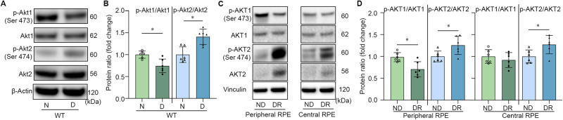
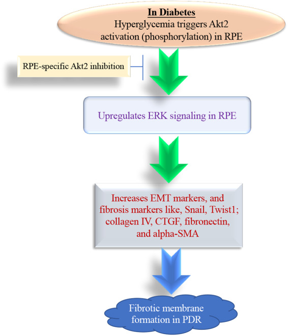
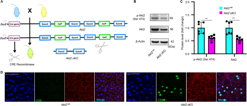
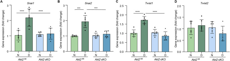
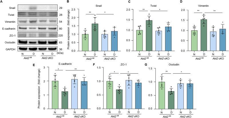
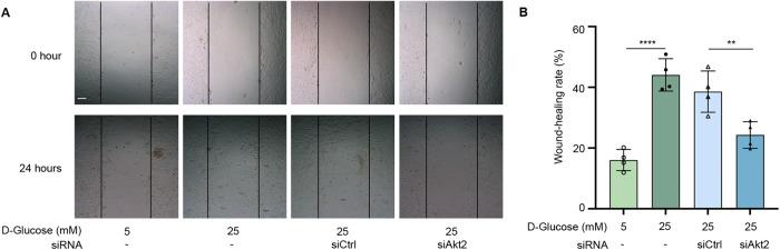
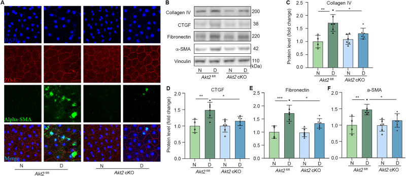
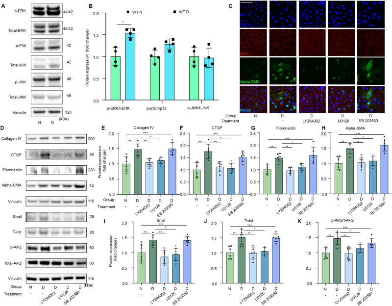
Diabetic Retinopathy (DR) is a complication of diabetes that causes blindness in adults. Retinal fibrosis is closely associated with developing proliferative diabetic retinopathy (PDR). Clinical studies have shown that fibrotic membranes exhibit uncontrolled growth in PDR and contribute to retinal detachment from RPE cells, ultimately leading to vision loss. While anti-VEGF agents and invasive laser treatments are the primary treatments for PDR, retinal fibrosis has received minimal attention as a potential target for therapeutic intervention. Therefore, to investigate the potential role of Akt2 in the diabetes-induced retinal fibrosis process, we generated RPE-specific Akt2 conditional knockout (cKO) mice and induced diabetes in these mice and Akt2 control mice by intraperitoneal injection of streptozotocin. After an 8-month duration of diabetes (10 months of age), the mice were euthanized and expression of tight junction proteins, epithelial-mesenchymal transition (EMT), and fibrosis markers were examined in the RPE. Diabetes induction in the floxed control mice decreased levels of the RPE tight junction protein ZO-1 and adherens junction proteins occludin and E-cadherin; these decreases were rescued in Akt2 cKO diabetic mice. Loss of Akt2 also inhibited diabetes-induced elevation of RNA and protein levels of the EMT markers Snail/Slug and Twist1 in the RPE as compared to Akt2 diabetic mice. We also found that in Akt2 cKO mice diabetes-induced increase of fibrosis markers, including collagen IV, Connective tissue growth factor (CTGF), fibronectin, and alpha-SMA was attenuated. Furthermore, we observed that high glucose-induced alterations in EMT and fibrosis markers in wild-type (WT) RPE explants were rescued in the presence of PI3K and ERK inhibitors, indicating diabetes-induced retinal fibrosis may be mediated via the PI3K/Akt2/ERK signaling, which could provide a novel target for DR therapy.
糖尿病性视网膜病变(DR)是糖尿病的一种并发症,可导致成年人失明。视网膜纤维化与增殖性糖尿病性视网膜病变(PDR)的发展密切相关。临床研究表明,纤维化膜在PDR中呈现不受控制的生长,并导致视网膜与视网膜色素上皮(RPE)细胞脱离,最终导致视力丧失。虽然抗血管内皮生长因子(VEGF)药物和侵入性激光治疗是PDR的主要治疗方法,但视网膜纤维化作为治疗干预的潜在靶点却很少受到关注。因此,为了研究Akt2在糖尿病诱导的视网膜纤维化过程中的潜在作用,我们构建了RPE特异性Akt2条件性敲除(cKO)小鼠,并通过腹腔注射链脲佐菌素在这些小鼠和Akt2对照小鼠中诱导糖尿病。糖尿病持续8个月(10月龄)后,对小鼠实施安乐死,并检测RPE中紧密连接蛋白、上皮-间质转化(EMT)和纤维化标志物的表达。在floxed对照小鼠中诱导糖尿病会降低RPE紧密连接蛋白ZO-1以及黏附连接蛋白闭合蛋白和E-钙黏蛋白的水平;而在Akt2 cKO糖尿病小鼠中这些降低的水平得到了恢复。与Akt2糖尿病小鼠相比,Akt2的缺失还抑制了糖尿病诱导的RPE中EMT标志物Snail/Slug和Twist1的RNA和蛋白质水平的升高。我们还发现,在Akt2 cKO小鼠中,糖尿病诱导的包括IV型胶原、结缔组织生长因子(CTGF)、纤连蛋白和α-平滑肌肌动蛋白(α-SMA)在内的纤维化标志物的增加有所减弱。此外,我们观察到在存在磷脂酰肌醇-3-激酶(PI3K)和细胞外信号调节激酶(ERK)抑制剂的情况下,野生型(WT)RPE外植体中高糖诱导的EMT和纤维化标志物的改变得到了恢复,这表明糖尿病诱导的视网膜纤维化可能是通过PI3K/Akt2/ERK信号传导介导的,这可能为DR治疗提供一个新的靶点。