• 文献检索
  • 文档翻译
  • 深度研究
  • 学术资讯
  • Suppr Zotero 插件Zotero 插件
  • 邀请有礼
  • 套餐&价格
  • 历史记录
应用&插件
Suppr Zotero 插件Zotero 插件浏览器插件Mac 客户端Windows 客户端微信小程序
定价
高级版会员购买积分包购买API积分包
服务
文献检索文档翻译深度研究API 文档MCP 服务
关于我们
关于 Suppr公司介绍联系我们用户协议隐私条款
关注我们

Suppr 超能文献

核心技术专利:CN118964589B侵权必究
粤ICP备2023148730 号-1Suppr @ 2026

文献检索

告别复杂PubMed语法,用中文像聊天一样搜索,搜遍4000万医学文献。AI智能推荐,让科研检索更轻松。

立即免费搜索

文件翻译

保留排版,准确专业,支持PDF/Word/PPT等文件格式,支持 12+语言互译。

免费翻译文档

深度研究

AI帮你快速写综述,25分钟生成高质量综述,智能提取关键信息,辅助科研写作。

立即免费体验

无论患者的容量状态如何,固有T2均可预测心肌炎症。

Native T2 Predicts Myocardial Inflammation Irrespective of a Patient's Volume Status.

作者信息

Wolter Jan Sebastian, Treiber Julia M, Fischer Selina, Fischer-Rasokat Ulrich, Kriechbaum Steffen D, Rieth Andreas, Weferling Maren, von Jeinsen Beatrice, Hain Andreas, Hamm Christian W, Keller Till, Rolf Andreas

机构信息

Kerckhoff Heart and Thorax Center, Department of Cardiology, Benekestrasse 2-8, 61231 Bad Nauheim, Germany.

German Center for Cardiovascular Research (DZHK), Rhine-Main Partner Site, 60590 Frankfurt am Main, Germany.

出版信息

Diagnostics (Basel). 2023 Jun 30;13(13):2240. doi: 10.3390/diagnostics13132240.

DOI:10.3390/diagnostics13132240
PMID:37443634
原文链接:https://pmc.ncbi.nlm.nih.gov/articles/PMC10340351/
Abstract

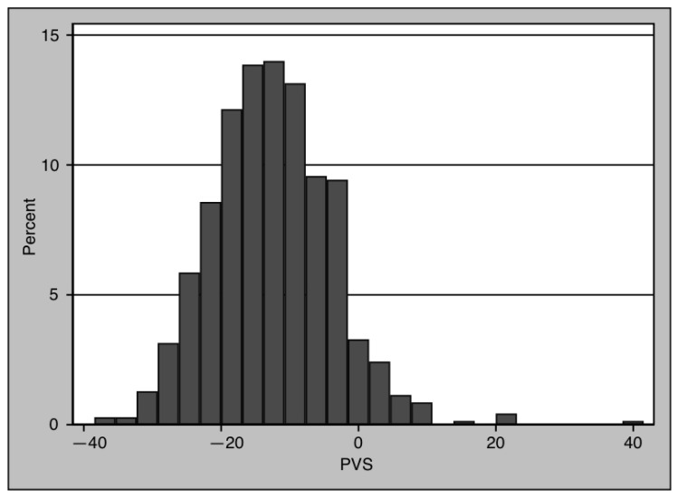
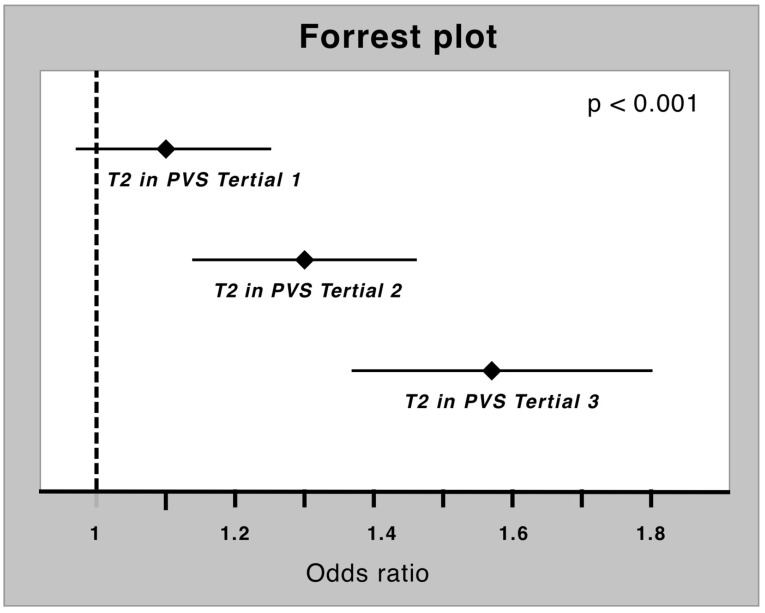
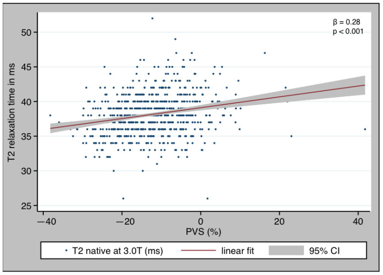
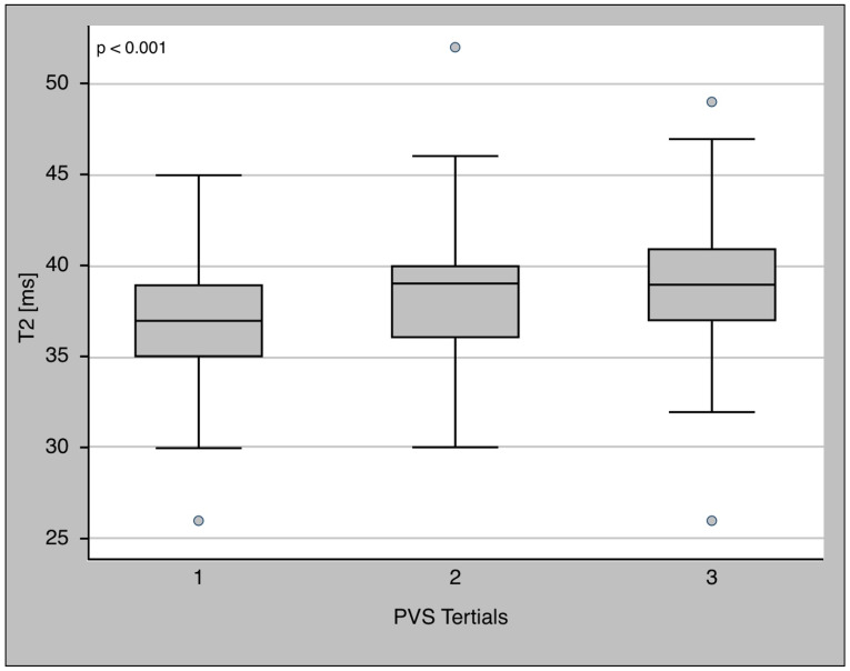
Myocardial inflammation and edema are major pathological features in myocarditis. Myocardial tissue water content and myocardial edema can be quantified via T2 mapping. Thus, cardiac magnetic resonance (CMR) is the noninvasive gold standard for diagnosing myocarditis. Several studies showed an impact of short-term volume changes on T2 relaxation time. Plasma volume status (PVS) is a good surrogate parameter to quantify a patient's volume status, and it is simple to use. The aim of this study was to determine the effect of PVS on the diagnostic value of T2 relaxation time in myocardial inflammation. Between April 2017 and December 2022, patients who were indicated for cardiac CMR were included in our prospective clinical registry. Patients with myocardial inflammation and those with unremarkable findings were analyzed in the present study. A blood sample was drawn, and PVS was calculated. Patients were separated into PVS tertiles to explore a possible nonlinear dose-response relationship. Logistic regression analysis was used to determine whether T2 is an independent predictor of myocardial inflammation. A total of 700 patients (47.43% female) were eligible for analysis. Of these, 551 patients were healthy (78.7%), while 149 (21.3%) showed signs of myocardial inflammation. The T2 relaxation time was elevated in patients with myocardial inflammation (40 ms [IQR 37-42 ms] vs. 38.0 ms [IQR 36-39 ms], < 0.001). PVS showed no difference between the groups (-12.94 [IQR -18.4--7.28] vs.-12.19 [IQR -18.93--5.87], = 0.384). T2 showed a clear dose-response relationship with PVS, with increasing T2 values along the PVS tertiles. In spite of this, T2 was found to be an independent marker of myocardial inflammation in logistic regression (OR T2 1.3 [95% CI 1.21-1.39], < 0.001), even after adjusting for PVS (OR T2 [adj. PVS] 1.31 [95% CI 1.22-1.40], < 0.001). Despite a dose-response relationship between T2 and the volume status, T2 was found to be an independent indicator of myocardial inflammation.

摘要

心肌炎症和水肿是心肌炎的主要病理特征。心肌组织含水量和心肌水肿可通过T2 mapping进行量化。因此,心脏磁共振成像(CMR)是诊断心肌炎的无创金标准。多项研究表明短期容量变化对T2弛豫时间有影响。血浆容量状态(PVS)是量化患者容量状态的一个良好替代参数,且使用简便。本研究的目的是确定PVS对心肌炎症中T2弛豫时间诊断价值的影响。2017年4月至2022年12月期间,被建议进行心脏CMR检查的患者被纳入我们的前瞻性临床登记研究。本研究分析了有心肌炎症的患者和检查结果无异常的患者。采集血样并计算PVS。将患者分为PVS三分位数组以探索可能的非线性剂量反应关系。采用逻辑回归分析来确定T2是否为心肌炎症的独立预测因子。共有700例患者(47.43%为女性)符合分析条件。其中,551例患者健康(78.7%),149例(21.3%)有心肌炎症迹象。有心肌炎症的患者T2弛豫时间升高(40 ms[四分位间距37 - 42 ms]对38.0 ms[四分位间距36 - 39 ms],<0.001)。两组间PVS无差异(-12.94[四分位间距-18.4 - -7.28]对-12.19[四分位间距-18.93 - -5.87],=0.384)。T2与PVS呈现明显的剂量反应关系,随着PVS三分位数升高T2值增加。尽管如此,在逻辑回归分析中发现T2是心肌炎症的独立标志物(T2的比值比为1.3[95%置信区间1.21 - 1.39],<0.001),即使在调整PVS后(调整PVS后的T2比值比为1.31[95%置信区间1.22 - 1.40],<0.001)。尽管T2与容量状态之间存在剂量反应关系,但T2被发现是心肌炎症的独立指标。

https://cdn.ncbi.nlm.nih.gov/pmc/blobs/ef6b/10340351/9dbbb005f09c/diagnostics-13-02240-g005.jpg
https://cdn.ncbi.nlm.nih.gov/pmc/blobs/ef6b/10340351/e6182fdfd3ae/diagnostics-13-02240-g001.jpg
https://cdn.ncbi.nlm.nih.gov/pmc/blobs/ef6b/10340351/dd762c0f1062/diagnostics-13-02240-g002.jpg
https://cdn.ncbi.nlm.nih.gov/pmc/blobs/ef6b/10340351/dbcda6d565eb/diagnostics-13-02240-g003.jpg
https://cdn.ncbi.nlm.nih.gov/pmc/blobs/ef6b/10340351/7efcc3f1d5bc/diagnostics-13-02240-g004.jpg
https://cdn.ncbi.nlm.nih.gov/pmc/blobs/ef6b/10340351/9dbbb005f09c/diagnostics-13-02240-g005.jpg

相似文献

1
Native T2 Predicts Myocardial Inflammation Irrespective of a Patient's Volume Status.无论患者的容量状态如何,固有T2均可预测心肌炎症。
Diagnostics (Basel). 2023 Jun 30;13(13):2240. doi: 10.3390/diagnostics13132240.
2
Native T1 is predictive of cardiovascular death/heart failure events and all-cause mortality irrespective of the patient's volume status.无论患者的容量状态如何,固有T1可预测心血管死亡/心力衰竭事件及全因死亡率。
Front Cardiovasc Med. 2023 Feb 14;10:1091334. doi: 10.3389/fcvm.2023.1091334. eCollection 2023.
3
Outcomes of Cardiovascular Magnetic Resonance Imaging in Patients Recently Recovered From Coronavirus Disease 2019 (COVID-19).COVID-19 患者近期康复后的心血管磁共振成像结果。
JAMA Cardiol. 2020 Nov 1;5(11):1265-1273. doi: 10.1001/jamacardio.2020.3557.
4
T(1) mapping for the diagnosis of acute myocarditis using CMR: comparison to T2-weighted and late gadolinium enhanced imaging.CMR 用于诊断急性心肌炎的 T(1) 映射:与 T2 加权和晚期钆增强成像的比较。
JACC Cardiovasc Imaging. 2013 Oct;6(10):1048-1058. doi: 10.1016/j.jcmg.2013.03.008. Epub 2013 Sep 4.
5
Comprehensive Cardiac Magnetic Resonance for Short-Term Follow-Up in Acute Myocarditis.急性心肌炎短期随访的综合心脏磁共振成像
J Am Heart Assoc. 2016 Jul 19;5(7):e003603. doi: 10.1161/JAHA.116.003603.
6
CMR in patients with severe myocarditis: diagnostic value of quantitative tissue markers including extracellular volume imaging.严重心肌炎患者的 CMR:包括细胞外容积成像在内的定量组织标志物的诊断价值。
JACC Cardiovasc Imaging. 2014 Jul;7(7):667-75. doi: 10.1016/j.jcmg.2014.02.005. Epub 2014 Jun 18.
7
Tissue characterization by T1 and T2 mapping cardiovascular magnetic resonance imaging to monitor myocardial inflammation in healing myocarditis.通过 T1 和 T2 映射心血管磁共振成像对心肌炎症进行组织特征描述以监测愈合性心肌炎。
Eur Heart J Cardiovasc Imaging. 2017 Jul 1;18(7):744-751. doi: 10.1093/ehjci/jex007.
8
Myocardial T1 and T2 Mapping by Magnetic Resonance in Patients With Immune Checkpoint Inhibitor-Associated Myocarditis.免疫检查点抑制剂相关心肌炎患者的磁共振心肌 T1 和 T2 mapping
J Am Coll Cardiol. 2021 Mar 30;77(12):1503-1516. doi: 10.1016/j.jacc.2021.01.050.
9
Cardiovascular Magnetic Resonance in Nonischemic Myocardial Inflammation: Expert Recommendations.非缺血性心肌炎症的心血管磁共振:专家建议。
J Am Coll Cardiol. 2018 Dec 18;72(24):3158-3176. doi: 10.1016/j.jacc.2018.09.072.
10
CMR-derived extracellular volume fraction (ECV) in asymptomatic heart transplant recipients: correlations with clinical features and myocardial edema.无症状心脏移植受者中磁共振成像衍生的细胞外容积分数(ECV):与临床特征及心肌水肿的相关性
Int J Cardiovasc Imaging. 2018 Dec;34(12):1959-1967. doi: 10.1007/s10554-018-1421-2. Epub 2018 Jul 28.

引用本文的文献

1
Effects of intraoperative open-lung strategy on cardiopulmonary function in frail elderly patients undergoing laparoscopic surgery: a randomized controlled trial.术中肺开放策略对老年体弱患者腹腔镜手术心肺功能的影响:一项随机对照试验
BMC Surg. 2025 Aug 6;25(1):346. doi: 10.1186/s12893-025-03115-0.

本文引用的文献

1
Native T1 is predictive of cardiovascular death/heart failure events and all-cause mortality irrespective of the patient's volume status.无论患者的容量状态如何,固有T1可预测心血管死亡/心力衰竭事件及全因死亡率。
Front Cardiovasc Med. 2023 Feb 14;10:1091334. doi: 10.3389/fcvm.2023.1091334. eCollection 2023.
2
Predictive value of overt and non-overt volume overload in patients with high- or low-gradient aortic stenosis undergoing transcatheter aortic valve implantation.有或无明显容量超负荷对接受经导管主动脉瓣植入术的高梯度或低梯度主动脉瓣狭窄患者的预测价值。
Cardiovasc Diagn Ther. 2021 Oct;11(5):1080-1092. doi: 10.21037/cdt-21-286.
3
Myocardial changes on 3T cardiovascular magnetic resonance imaging in response to haemodialysis with fluid removal.
3T 心血管磁共振成像中血液透析去除液体后心肌变化。
J Cardiovasc Magn Reson. 2021 Nov 11;23(1):125. doi: 10.1186/s12968-021-00822-4.
4
Myocarditis and inflammatory cardiomyopathy: current evidence and future directions.心肌炎和炎性心肌病:当前的证据和未来的方向。
Nat Rev Cardiol. 2021 Mar;18(3):169-193. doi: 10.1038/s41569-020-00435-x. Epub 2020 Oct 12.
5
Influence of hydration status on cardiovascular magnetic resonance myocardial T1 and T2 relaxation time assessment: an intraindividual study in healthy subjects.探讨水合状态对心血管磁共振心肌 T1 和 T2 弛豫时间评估的影响:健康受试者的个体内研究。
J Cardiovasc Magn Reson. 2020 Sep 7;22(1):63. doi: 10.1186/s12968-020-00661-9.
6
Ventricular Arrhythmias and Sudden Cardiac Death in Lymphocytic Myocarditis.淋巴细胞性心肌炎中的室性心律失常与心源性猝死
J Am Coll Cardiol. 2020 Mar 10;75(9):1058-1060. doi: 10.1016/j.jacc.2020.01.032.
7
Sudden cardiac death in patients with myocarditis: Evaluation, risk stratification, and management.心肌炎患者的心脏性猝死:评估、危险分层与管理。
Am Heart J. 2020 Feb;220:29-40. doi: 10.1016/j.ahj.2019.08.007. Epub 2019 Aug 15.
8
Management of Myocarditis-Related Cardiomyopathy in Adults.成人心肌炎相关性心肌病的管理。
Circ Res. 2019 May 24;124(11):1568-1583. doi: 10.1161/CIRCRESAHA.118.313578.
9
Acute changes in cardiac structural and tissue characterisation parameters following haemodialysis measured using cardiovascular magnetic resonance.应用心血管磁共振测量血液透析后心脏结构和组织特征参数的急性变化。
Sci Rep. 2019 Feb 4;9(1):1388. doi: 10.1038/s41598-018-37845-4.
10
Cardiovascular Magnetic Resonance in Nonischemic Myocardial Inflammation: Expert Recommendations.非缺血性心肌炎症的心血管磁共振:专家建议。
J Am Coll Cardiol. 2018 Dec 18;72(24):3158-3176. doi: 10.1016/j.jacc.2018.09.072.