• 文献检索
  • 文档翻译
  • 深度研究
  • 学术资讯
  • Suppr Zotero 插件Zotero 插件
  • 邀请有礼
  • 套餐&价格
  • 历史记录
应用&插件
Suppr Zotero 插件Zotero 插件浏览器插件Mac 客户端Windows 客户端微信小程序
定价
高级版会员购买积分包购买API积分包
服务
文献检索文档翻译深度研究API 文档MCP 服务
关于我们
关于 Suppr公司介绍联系我们用户协议隐私条款
关注我们

Suppr 超能文献

核心技术专利:CN118964589B侵权必究
粤ICP备2023148730 号-1Suppr @ 2026

文献检索

告别复杂PubMed语法,用中文像聊天一样搜索,搜遍4000万医学文献。AI智能推荐,让科研检索更轻松。

立即免费搜索

文件翻译

保留排版,准确专业,支持PDF/Word/PPT等文件格式,支持 12+语言互译。

免费翻译文档

深度研究

AI帮你快速写综述,25分钟生成高质量综述,智能提取关键信息,辅助科研写作。

立即免费体验

使用连续块面扫描电子显微镜(SBF-SEM)对人类细胞有丝分裂保真度进行3D超微结构可视化

3D Ultrastructural Visualization of Mitosis Fidelity in Human Cells Using Serial Block Face Scanning Electron Microscopy (SBF-SEM).

作者信息

Ferrandiz Nuria, Royle Stephen J

机构信息

Centre for Mechanochemical Cell Biology and Division of Biomedical Sciences, Warwick Medical School, University of Warwick, Gibbet Hill Road, Coventry, CV4 7AL, UK.

出版信息

Bio Protoc. 2023 Jul 5;13(13):e4708. doi: 10.21769/BioProtoc.4708.

DOI:10.21769/BioProtoc.4708
PMID:37449034
原文链接:https://pmc.ncbi.nlm.nih.gov/articles/PMC10336565/
Abstract

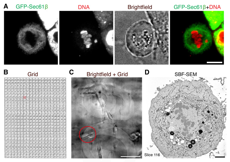
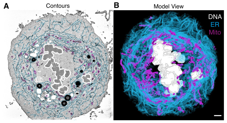
Errors in chromosome segregation during mitosis lead to chromosome instability, resulting in an unbalanced number of chromosomes in the daughter cells. Light microscopy has been used extensively to study chromosome missegregation by visualizing errors of the mitotic spindle. However, less attention has been paid to understanding spindle function in the broader context of intracellular structures and organelles during mitosis. Here, we outline a protocol to visualize chromosomes and endomembranes in mitosis, combining light microscopy and 3D volume electron microscopy, serial block-face scanning electron microscopy (SBF-SEM). SBF-SEM provides high-resolution imaging of large volumes and subcellular structures, followed by image analysis and 3D reconstruction. This protocol allows scientists to visualize the whole subcellular context of the spindle during mitosis.

摘要

有丝分裂期间染色体分离错误会导致染色体不稳定,从而使子细胞中的染色体数量失衡。光学显微镜已被广泛用于通过观察有丝分裂纺锤体的错误来研究染色体错分离。然而,在有丝分裂期间细胞内结构和细胞器的更广泛背景下,对纺锤体功能的理解却较少受到关注。在这里,我们概述了一种在有丝分裂中可视化染色体和内膜的方案,该方案结合了光学显微镜和三维体积电子显微镜——连续块面扫描电子显微镜(SBF-SEM)。SBF-SEM可对大体积和亚细胞结构进行高分辨率成像,随后进行图像分析和三维重建。该方案使科学家能够在有丝分裂期间可视化纺锤体的整个亚细胞背景。

https://cdn.ncbi.nlm.nih.gov/pmc/blobs/4d4a/10336565/ba5c7096d2e6/BioProtoc-13-13-4708-g002.jpg
https://cdn.ncbi.nlm.nih.gov/pmc/blobs/4d4a/10336565/a120546ebf17/BioProtoc-13-13-4708-g001.jpg
https://cdn.ncbi.nlm.nih.gov/pmc/blobs/4d4a/10336565/ba5c7096d2e6/BioProtoc-13-13-4708-g002.jpg
https://cdn.ncbi.nlm.nih.gov/pmc/blobs/4d4a/10336565/a120546ebf17/BioProtoc-13-13-4708-g001.jpg
https://cdn.ncbi.nlm.nih.gov/pmc/blobs/4d4a/10336565/ba5c7096d2e6/BioProtoc-13-13-4708-g002.jpg

相似文献

1
3D Ultrastructural Visualization of Mitosis Fidelity in Human Cells Using Serial Block Face Scanning Electron Microscopy (SBF-SEM).使用连续块面扫描电子显微镜(SBF-SEM)对人类细胞有丝分裂保真度进行3D超微结构可视化
Bio Protoc. 2023 Jul 5;13(13):e4708. doi: 10.21769/BioProtoc.4708.
2
Correlating light microscopy with serial block face scanning electron microscopy to study mitotic spindle architecture.将光学显微镜与连续块面扫描电子显微镜相结合以研究有丝分裂纺锤体结构。
Methods Cell Biol. 2018;145:29-43. doi: 10.1016/bs.mcb.2018.03.010.
3
Serial Block Face-Scanning Electron Microscopy as a Burgeoning Technology.序列块面扫描电子显微镜技术的兴起。
Adv Biol (Weinh). 2023 Aug;7(8):e2300139. doi: 10.1002/adbi.202300139. Epub 2023 May 28.
4
Comparison of 3D cellular imaging techniques based on scanned electron probes: Serial block face SEM vs. Axial bright-field STEM tomography.基于扫描电子探针的 3D 细胞成像技术比较:连续块面扫描电镜与轴向明场扫描透射电镜断层成像。
J Struct Biol. 2018 Jun;202(3):216-228. doi: 10.1016/j.jsb.2018.01.012. Epub 2018 Feb 1.
5
A workflow for 3D-CLEM investigating liver tissue.用于研究肝组织的 3D-CLEM 工作流程。
J Microsc. 2021 Mar;281(3):231-242. doi: 10.1111/jmi.12967. Epub 2020 Oct 27.
6
Combining serial block face and focused ion beam scanning electron microscopy for 3D studies of rare events.结合连续块面和聚焦离子束扫描电子显微镜用于罕见事件的三维研究。
Methods Cell Biol. 2019;152:87-101. doi: 10.1016/bs.mcb.2019.03.014. Epub 2019 May 7.
7
Protocols for Generating Surfaces and Measuring 3D Organelle Morphology Using Amira.使用 Amira 生成表面和测量 3D 细胞器形态的方案。
Cells. 2021 Dec 27;11(1):65. doi: 10.3390/cells11010065.
8
Development of protocols for the first serial block-face scanning electron microscopy (SBF SEM) studies of bone tissue.制定用于骨组织的第一代连续块面扫描电子显微镜 (SBF SEM) 研究的方案。
Bone. 2020 Feb;131:115107. doi: 10.1016/j.bone.2019.115107. Epub 2019 Oct 24.
9
Endomembranes promote chromosome missegregation by ensheathing misaligned chromosomes.内质网通过包裹未对齐的染色体促进染色体错误分离。
J Cell Biol. 2022 Jun 6;221(6). doi: 10.1083/jcb.202203021. Epub 2022 Apr 29.
10
Mesoscale morphology at nanoscale resolution: serial block-face scanning electron microscopy reveals fine 3D detail of a novel silk spinneret system in a tube-building tanaid crustacean.纳米级分辨率下的中尺度形态:连续块面扫描电子显微镜揭示了一种管栖等足甲壳动物新型吐丝器系统的精细三维细节。
Front Zool. 2016 Mar 22;13:14. doi: 10.1186/s12983-016-0146-0. eCollection 2016.

引用本文的文献

1
Nondisruptive inducible labeling of ER-membrane contact sites using the Lamin B receptor.使用核纤层蛋白B受体对内质网-膜接触位点进行非破坏性诱导标记
PLoS Biol. 2025 Jul 10;23(7):e3003249. doi: 10.1371/journal.pbio.3003249. eCollection 2025 Jul.
2
Sample preparation and data collection for serial block face scanning electron microscopy of mammalian cell monolayers.哺乳动物细胞单层的连续块面扫描电子显微镜的样品制备和数据收集。
PLoS One. 2024 Aug 9;19(8):e0301284. doi: 10.1371/journal.pone.0301284. eCollection 2024.

本文引用的文献

1
Endomembranes promote chromosome missegregation by ensheathing misaligned chromosomes.内质网通过包裹未对齐的染色体促进染色体错误分离。
J Cell Biol. 2022 Jun 6;221(6). doi: 10.1083/jcb.202203021. Epub 2022 Apr 29.
2
Microtubule organization within mitotic spindles revealed by serial block face scanning electron microscopy and image analysis.通过连续块面扫描电子显微镜和图像分析揭示有丝分裂纺锤体内的微管组织。
J Cell Sci. 2017 May 15;130(10):1845-1855. doi: 10.1242/jcs.203877. Epub 2017 Apr 7.
3
Cellular Reorganization during Mitotic Entry.
细胞在有丝分裂进入时的重新组织。
Trends Cell Biol. 2017 Jan;27(1):26-41. doi: 10.1016/j.tcb.2016.07.004. Epub 2016 Aug 12.
4
Microscopy Image Browser: A Platform for Segmentation and Analysis of Multidimensional Datasets.显微镜图像浏览器:一个用于多维数据集分割与分析的平台。
PLoS Biol. 2016 Jan 4;14(1):e1002340. doi: 10.1371/journal.pbio.1002340. eCollection 2016 Jan.
5
Fiji: an open-source platform for biological-image analysis.斐济:一个用于生物影像分析的开源平台。
Nat Methods. 2012 Jun 28;9(7):676-82. doi: 10.1038/nmeth.2019.
6
Progressive sheet-to-tubule transformation is a general mechanism for endoplasmic reticulum partitioning in dividing mammalian cells.板状到管状的渐进转化是哺乳动物细胞分裂中内质网分区的一般机制。
Mol Biol Cell. 2012 Jul;23(13):2424-32. doi: 10.1091/mbc.E10-12-0950. Epub 2012 May 9.
7
Cisternal organization of the endoplasmic reticulum during mitosis.有丝分裂过程中内质网的池状组织。
Mol Biol Cell. 2009 Aug;20(15):3471-80. doi: 10.1091/mbc.e09-04-0327. Epub 2009 Jun 3.
8
Endoplasmic reticulum remains continuous and undergoes sheet-to-tubule transformation during cell division in mammalian cells.内质网保持连续,并在哺乳动物细胞分裂过程中经历从片状到管状的转变。
J Cell Biol. 2007 Dec 3;179(5):895-909. doi: 10.1083/jcb.200705112.
9
Computer visualization of three-dimensional image data using IMOD.使用IMOD对三维图像数据进行计算机可视化。
J Struct Biol. 1996 Jan-Feb;116(1):71-6. doi: 10.1006/jsbi.1996.0013.