Carnegie Mellon University, Neuroscience Institute, Pittsburgh, Pennsylvania, United States.
Carnegie Mellon University, Department of Biomedical Engineering, Pittsburgh, Pennsylvania, United States.
J Biomed Opt. 2023 Jul;28(7):075001. doi: 10.1117/1.JBO.28.7.075001. Epub 2023 Jul 13.
Using functional near-infrared spectroscopy (fNIRS) in bottlenose dolphins () could help to understand how echolocating animals perceive their environment and how they focus on specific auditory objects, such as fish, in noisy marine settings.
To test the feasibility of near-infrared spectroscopy (NIRS) in medium-sized marine mammals, such as dolphins, we modeled the light propagation with computational tools to determine the wavelengths, optode locations, and separation distances that maximize sensitivity to brain tissue.
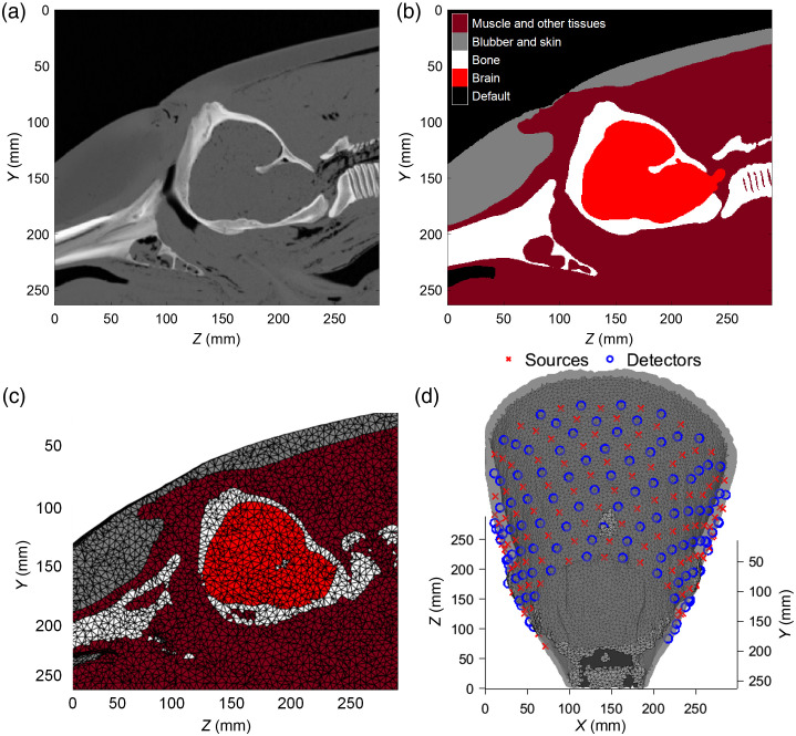
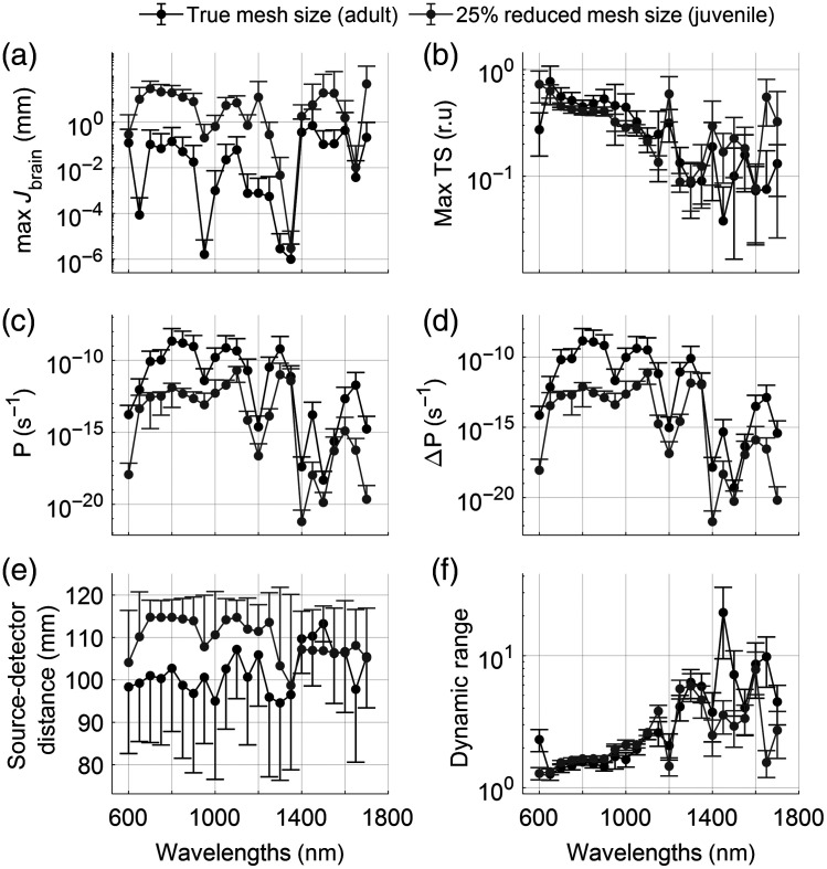
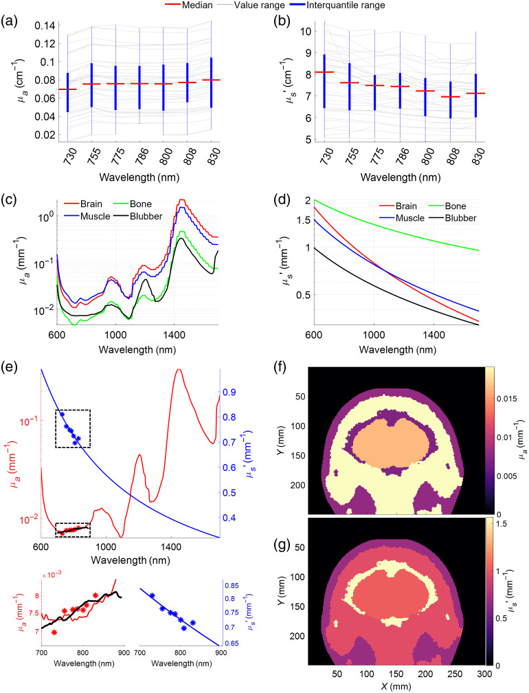
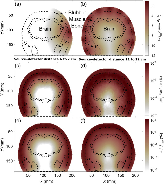
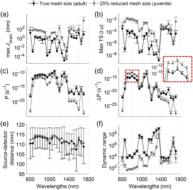
Using frequency-domain NIRS, we measured the absorption and reduced scattering coefficient of dolphin sculp. We assigned muscle, bone, and brain optical properties from the literature and modeled light propagation in a spatially accurate and biologically relevant model of a dolphin head, using finite-element modeling. We assessed tissue sensitivities for a range of wavelengths (600 to 1700 nm), source-detector distances (50 to 120 mm), and animal sizes (juvenile model 25% smaller than adult).
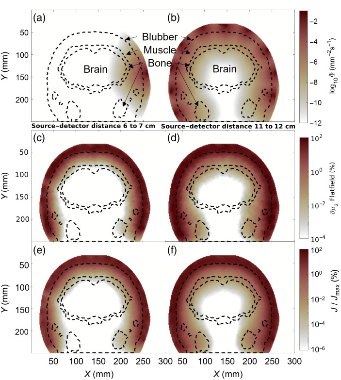
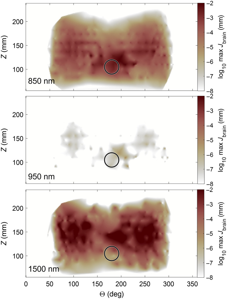
We found that the wavelengths most suitable for imaging the brain fell into two ranges: 700 to 900 nm and 1100 to 1150 nm. The optimal location for brain sensing positioned the center point between source and detector 30 to 50 mm caudal of the blowhole and at an angle 45 deg to 90 deg lateral off the midsagittal plane. Brain tissue sensitivity comparable to human measurements appears achievable only for smaller animals, such as juvenile bottlenose dolphins or smaller species of cetaceans, such as porpoises, or with source-detector separations in adult dolphins.
Brain measurements in juvenile or subadult dolphins, or smaller dolphin species, may be possible using specialized fNIRS devices that support optode separations of . We speculate that many measurement repetitions will be required to overcome hemodynamic signals originating predominantly from the muscle layer above the skull. NIRS measurements of muscle tissue are feasible today with source-detector separations of 50 mm, or even less.
在宽吻海豚()中使用功能近红外光谱(fNIRS)可以帮助我们理解回声定位动物如何感知其环境,以及它们如何在嘈杂的海洋环境中专注于特定的听觉物体,例如鱼类。
为了测试近红外光谱(NIRS)在中型海洋哺乳动物(如海豚)中的可行性,我们使用计算工具模拟光的传播,以确定最大限度提高脑组织灵敏度的波长、光纤位置和分离距离。
使用频域 NIRS,我们测量了海豚颅骨的吸收和散射系数减少量。我们从文献中分配了肌肉、骨骼和大脑的光学特性,并使用有限元建模在海豚头部的空间准确和生物学相关模型中模拟光的传播。我们评估了一系列波长(600 至 1700nm)、源-探测器距离(50 至 120mm)和动物大小(比成年模型小 25%的幼体模型)的组织灵敏度。
我们发现,最适合大脑成像的波长分为两个范围:700 至 900nm 和 1100 至 1150nm。用于脑传感的最佳位置是将源和探测器之间的中心点放置在呼吸孔后方 30 至 50mm 处,并且与正中矢状面成 45 度至 90 度的角度。只有对于较小的动物,如幼年宽吻海豚或较小的鲸类物种,如海豚,或者源-探测器分离距离在成年海豚中,才能实现与人类测量相当的脑组织灵敏度。
使用支持光纤分离的专用 fNIRS 设备,可能可以对幼年或亚成年海豚或较小的海豚物种进行脑部测量。我们推测,需要进行许多测量重复才能克服主要源自颅骨上方肌肉层的血液动力学信号。使用源-探测器分离距离为 50mm 或更小的距离,今天已经可以进行肌肉组织的 NIRS 测量。