University of Health Sciences, Tepecik Education and Research Hospital Center, Department of Radiology - Izmir, Turkey.
University of Health Sciences, Tepecik Education and Research Hospital Center, Department of Gynaecologic Oncology - Izmir, Turkey.
Rev Assoc Med Bras (1992). 2023 Jul 17;69(7):e20230110. doi: 10.1590/1806-9282.20230110. eCollection 2023.
This study was carried out to investigate the differentiation of mucinous borderline ovarian tumor from mucinous ovarian carcinoma using magnetic resonance imaging.
We evaluated 77 women patients who underwent abdominal magnetic resonance imaging due to pelvic mass. magnetic resonance imaging was reviewed by an experienced radiologist. A total of 70 women patients were included in the study. The magnetic resonance imaging features were retrospectively evaluated and compared between the two pathologies.
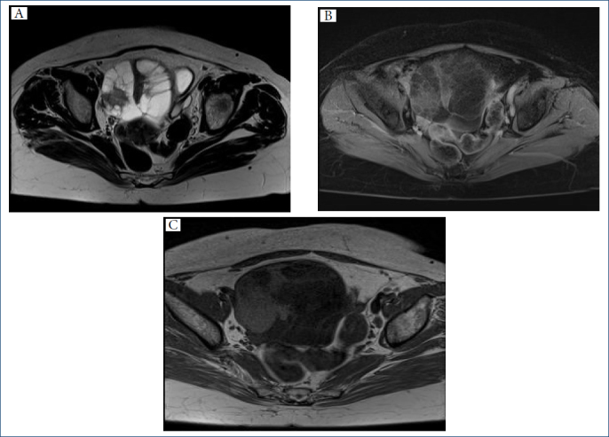
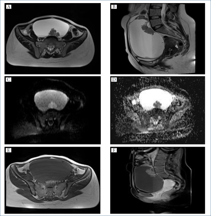
There was no difference between the two groups in terms of maximum tumor size. Age at diagnosis was 56.29±11.92 in the mucinous ovarian carcinoma group and 44.74±13.60 in the mucinous borderline ovarian tumor group (p<0.05). A significant difference was found between the two groups, and it was observed that mucinous borderline ovarian tumors appeared in the younger age group compared to mucinous ovarian carcinomas. Presence of ascites, peritoneal dissemination, lymphadenopathy, and mural nodules was found significantly more frequently in mucinous ovarian carcinomas than in mucinous borderline ovarian tumors. Honeycomb appearance was found more frequently in mucinous borderline ovarian tumor patients than in mucinous ovarian carcinoma patients.
magnetic resonance imaging findings of these two pathologies overlapped considerably. Compared with mucinous borderline ovarian tumors, mucinous ovarian carcinomas frequently had mural nodules larger than 5 mm, larger tumor size, peritoneal dissemination, and abnormal ascites.
本研究旨在通过磁共振成像(MRI)探讨黏液性交界性卵巢肿瘤与黏液性卵巢癌的鉴别诊断。
我们评估了 77 例因盆腔肿块而行腹部 MRI 检查的女性患者。由一位经验丰富的放射科医生对 MRI 进行了评估。共有 70 例女性患者纳入本研究。回顾性评估并比较了两种病理类型的 MRI 特征。
两组患者的最大肿瘤大小无差异。黏液性卵巢癌组的诊断年龄为 56.29±11.92 岁,黏液性交界性卵巢肿瘤组为 44.74±13.60 岁(p<0.05)。两组之间存在显著差异,黏液性交界性卵巢肿瘤组的发病年龄明显低于黏液性卵巢癌组。黏液性卵巢癌组中腹水、腹膜播散、淋巴结病和壁结节的存在明显多于黏液性交界性卵巢肿瘤组。黏液性交界性卵巢肿瘤组中出现蜂窝状外观的患者多于黏液性卵巢癌组。
这两种病变的 MRI 表现有很大的重叠。与黏液性交界性卵巢肿瘤相比,黏液性卵巢癌常伴有大于 5mm 的壁结节、更大的肿瘤大小、腹膜播散和异常腹水。