Meinig School of Biomedical Engineering, Cornell University, Ithaca, NY 14850, USA.
Department of Radiology, Weill Cornell Medicine, New York, NY 10065, USA.
Tomography. 2023 Jul 12;9(4):1341-1355. doi: 10.3390/tomography9040107.
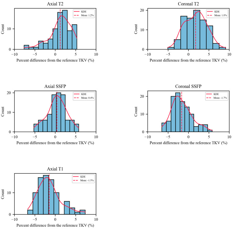
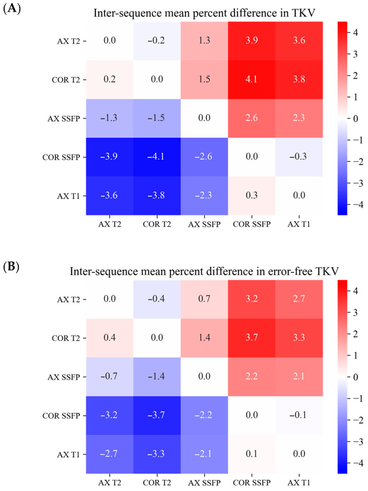
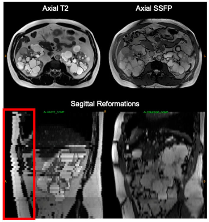
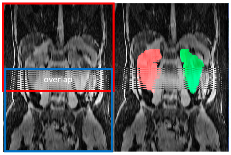
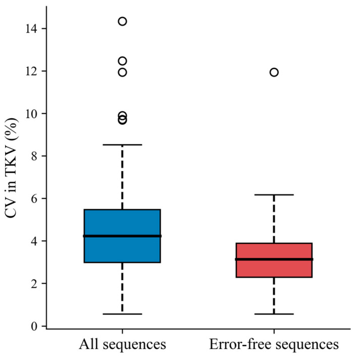
Total kidney volume measured on MRI is an important biomarker for assessing the progression of autosomal dominant polycystic kidney disease and response to treatment. However, we have noticed that there can be substantial differences in the kidney volume measurements obtained from the various pulse sequences commonly included in an MRI exam. Here we examine kidney volume measurement variability among five commonly acquired MRI pulse sequences in abdominal MRI exams in 105 patients with ADPKD. Right and left kidney volumes were independently measured by three expert observers using model-assisted segmentation for axial T2, coronal T2, axial single-shot fast spin echo (SSFP), coronal SSFP, and axial 3D T1 images obtained on a single MRI from ADPKD patients. Outlier measurements were analyzed for data acquisition errors. Most of the outlier values (88%) were due to breathing during scanning causing slice misregistration with gaps or duplication of imaging slices ( = 35), slice misregistration from using multiple breath holds during acquisition ( = 25), composing of two overlapping acquisitions ( = 17), or kidneys not entirely within the field of view ( = 4). After excluding outlier measurements, the coefficient of variation among the five measurements decreased from 4.6% pre to 3.2%. Compared to the average of all sequences without errors, TKV measured on axial and coronal T2 weighted imaging were 1.2% and 1.8% greater, axial SSFP was 0.4% greater, coronal SSFP was 1.7% lower and axial T1 was 1.5% lower than the mean, indicating intrinsic measurement biases related to the different MRI contrast mechanisms. In conclusion, MRI data acquisition errors are common but can be identified using outlier analysis and excluded to improve organ volume measurement consistency. Bias toward larger volume measurements on T2 sequences and smaller volumes on axial T1 sequences can also be mitigated by averaging data from all error-free sequences acquired.
MRI 测量的全肾体积是评估常染色体显性多囊肾病(autosomal dominant polycystic kidney disease,ADPKD)进展和治疗反应的重要生物标志物。然而,我们注意到,在 MRI 检查中通常包含的各种脉冲序列中,获得的肾体积测量值可能存在很大差异。在这里,我们检查了 105 例 ADPKD 患者腹部 MRI 检查中 5 种常用 MRI 脉冲序列的肾体积测量变异性。三位专家观察者分别使用模型辅助分割对轴向 T2、冠状 T2、轴向单次激发快速自旋回波(single-shot fast spin echo,SSFP)、冠状 SSFP 和轴向 3D T1 图像进行独立测量。对离群值测量进行了数据采集误差分析。大多数离群值(88%)是由于扫描过程中的呼吸导致成像层面错位,出现间隙或重复成像层面( = 35),采集过程中使用多次屏气导致层面错位( = 25),两个重叠采集( = 17),或肾脏不完全在视野内( = 4)。排除离群值测量后,五种测量方法之间的变异系数从之前的 4.6%降至 3.2%。与无误差的所有序列平均值相比,轴向和冠状 T2 加权成像的 TKV 分别增加了 1.2%和 1.8%,轴向 SSFP 增加了 0.4%,冠状 SSFP 减少了 1.7%,轴向 T1 减少了 1.5%,这表明与不同 MRI 对比机制相关的固有测量偏差。总之,MRI 数据采集误差很常见,但可以通过离群值分析识别并排除,以提高器官体积测量的一致性。通过平均所有无误差序列的数据,也可以减轻 T2 序列上的体积测量偏差和轴向 T1 序列上的体积测量偏差。