Department of Radiology, Weill Cornell Medicine, Cornell University, New York, NY 10065, USA.
The Rogosin Institute and Department of Medicine Weill Cornell Medicine, Cornell University, New York, NY 10065, USA.
Tomography. 2022 Jul 13;8(4):1804-1819. doi: 10.3390/tomography8040152.
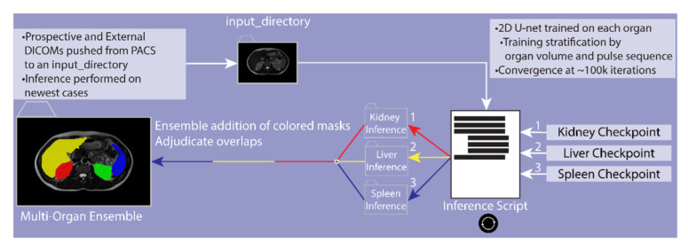
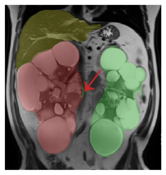
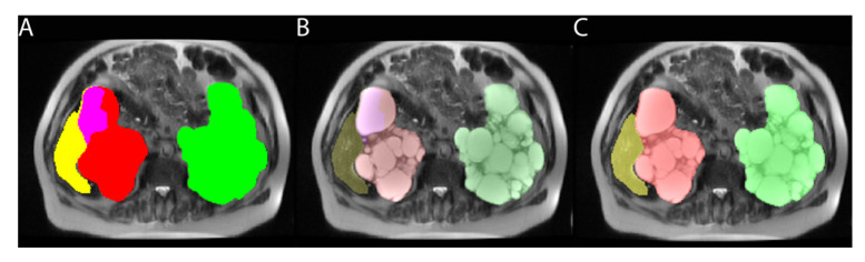
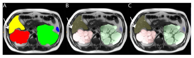
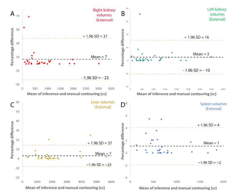
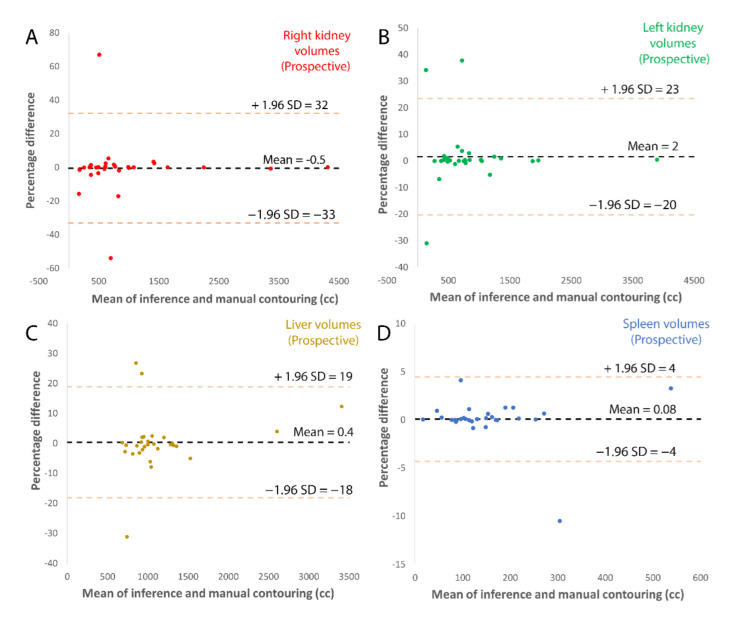
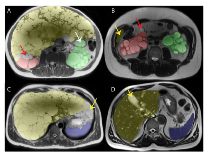
Organ volume measurements are a key metric for managing ADPKD (the most common inherited renal disease). However, measuring organ volumes is tedious and involves manually contouring organ outlines on multiple cross-sectional MRI or CT images. The automation of kidney contouring using deep learning has been proposed, as it has small errors compared to manual contouring. Here, a deployed open-source deep learning ADPKD kidney segmentation pipeline is extended to also measure liver and spleen volumes, which are also important. This 2D U-net deep learning approach was developed with radiologist labeled T2-weighted images from 215 ADPKD subjects (70% training = 151, 30% validation = 64). Additional ADPKD subjects were utilized for prospective (n = 30) and external (n = 30) validations for a total of 275 subjects. Image cropping previously optimized for kidneys was included in training but removed for the validation and inference to accommodate the liver which is closer to the image border. An effective algorithm was developed to adjudicate overlap voxels that are labeled as more than one organ. Left kidney, right kidney, liver and spleen labels had average errors of 3%, 7%, 3%, and 1%, respectively, on external validation and 5%, 6%, 5%, and 1% on prospective validation. Dice scores also showed that the deep learning model was close to the radiologist contouring, measuring 0.98, 0.96, 0.97 and 0.96 on external validation and 0.96, 0.96, 0.96 and 0.95 on prospective validation for left kidney, right kidney, liver and spleen, respectively. The time required for manual correction of deep learning segmentation errors was only 19:17 min compared to 33:04 min for manual segmentations, a 42% time saving ( = 0.004). Standard deviation of model assisted segmentations was reduced to 7, 5, 11, 5 mL for right kidney, left kidney, liver and spleen respectively from 14, 10, 55 and 14 mL for manual segmentations. Thus, deep learning reduces the radiologist time required to perform multiorgan segmentations in ADPKD and reduces measurement variability.
器官体积测量是管理常染色体显性多囊肾病(最常见的遗传性肾脏疾病)的关键指标。然而,测量器官体积是一项繁琐的工作,需要在多个磁共振成像或 CT 图像上手动描绘器官轮廓。使用深度学习自动化肾脏轮廓描绘已被提出,因为与手动描绘相比,它的误差较小。在这里,扩展了已部署的开源深度学习 ADPKD 肾脏分割管道,以测量肝脏和脾脏的体积,这也很重要。这种二维 U 形网络深度学习方法是使用来自 215 名 ADPKD 患者的放射科医生标记的 T2 加权图像开发的(70%的训练= 151,30%的验证= 64)。另外,还对 30 名前瞻性(n=30)和 30 名外部(n=30)验证患者进行了验证,总共有 275 名患者。之前针对肾脏进行了优化的图像裁剪包括在训练中,但在验证和推断中删除,以适应更靠近图像边界的肝脏。开发了一种有效的算法来解决被标记为多个器官的重叠体素。在外部验证中,左肾、右肾、肝和脾标签的平均误差分别为 3%、7%、3%和 1%,前瞻性验证中分别为 5%、6%、5%和 1%。Dice 评分还表明,深度学习模型与放射科医生的轮廓非常接近,在外部验证中,左肾、右肾、肝和脾的 Dice 评分为 0.98、0.96、0.97 和 0.96,在前瞻性验证中,Dice 评分为 0.96、0.96、0.96 和 0.95。与手动分割相比,深度学习分割错误的手动校正所需的时间仅为 19:17 分钟,节省了 42%的时间(=0.004)。从手动分割的 14、10、55 和 14 毫升,将模型辅助分割的标准偏差分别降低到右肾、左肾、肝和脾的 7、5、11 和 5 毫升。因此,深度学习减少了放射科医生在 ADPKD 中进行多器官分割所需的时间,并降低了测量的可变性。