• 文献检索
  • 文档翻译
  • 深度研究
  • 学术资讯
  • Suppr Zotero 插件Zotero 插件
  • 邀请有礼
  • 套餐&价格
  • 历史记录
应用&插件
Suppr Zotero 插件Zotero 插件浏览器插件Mac 客户端Windows 客户端微信小程序
定价
高级版会员购买积分包购买API积分包
服务
文献检索文档翻译深度研究API 文档MCP 服务
关于我们
关于 Suppr公司介绍联系我们用户协议隐私条款
关注我们

Suppr 超能文献

核心技术专利:CN118964589B侵权必究
粤ICP备2023148730 号-1Suppr @ 2026

文献检索

告别复杂PubMed语法,用中文像聊天一样搜索,搜遍4000万医学文献。AI智能推荐,让科研检索更轻松。

立即免费搜索

文件翻译

保留排版,准确专业,支持PDF/Word/PPT等文件格式,支持 12+语言互译。

免费翻译文档

深度研究

AI帮你快速写综述,25分钟生成高质量综述,智能提取关键信息,辅助科研写作。

立即免费体验

经胸超声心动图评估主动脉瓣狭窄的陷阱与技巧

Pitfalls and Tips in the Assessment of Aortic Stenosis by Transthoracic Echocardiography.

作者信息

Canciello Grazia, Pate Shabnam, Sannino Anna, Borrelli Felice, Todde Gaetano, Grayburn Paul, Losi Maria-Angela, Esposito Giovanni

机构信息

Department of Advanced Biomedical Sciences, Federico II University, 80131 Naples, Italy.

Division of Cardiology, Baylor Scott & White Research Institute, Plano, TX 75204, USA.

出版信息

Diagnostics (Basel). 2023 Jul 19;13(14):2414. doi: 10.3390/diagnostics13142414.

DOI:10.3390/diagnostics13142414
PMID:37510158
原文链接:https://pmc.ncbi.nlm.nih.gov/articles/PMC10377988/
Abstract

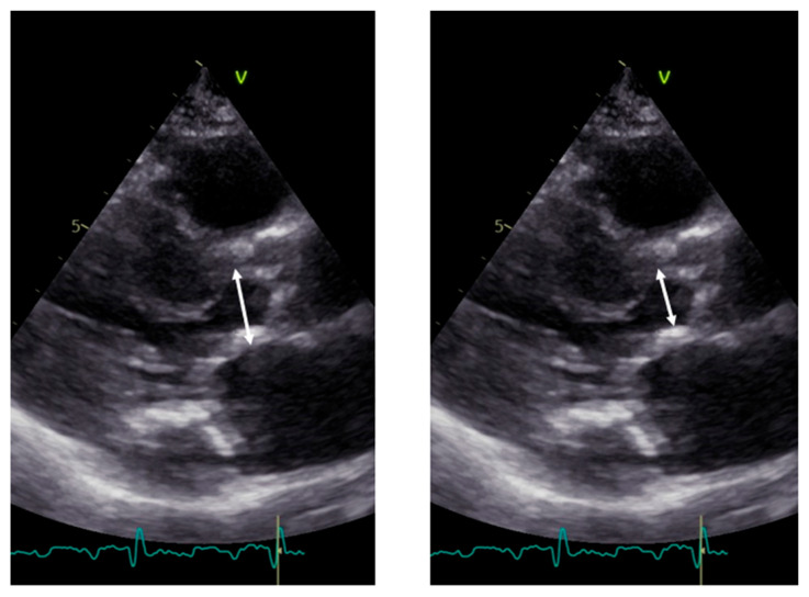
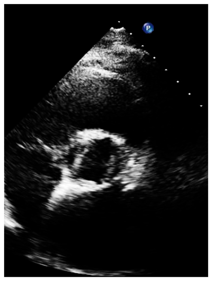
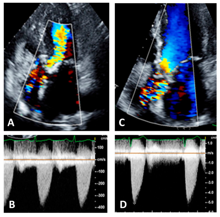
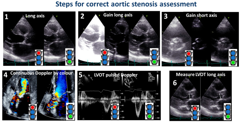
Aortic stenosis (AS) is a valvular heart disease that significantly contributes to cardiovascular morbidity and mortality worldwide. The condition is characterized by calcification and thickening of the aortic valve leaflets, resulting in a narrowed orifice and increased pressure gradient across the valve. AS typically progresses from a subclinical phase known as aortic sclerosis, where valve calcification occurs without a transvalvular gradient, to a more advanced stage marked by a triad of symptoms: heart failure, syncope, and angina. Echocardiography plays a crucial role in the diagnosis and evaluation of AS, serving as the primary non-invasive imaging modality. However, to minimize misdiagnoses, it is crucial to adhere to a standardized protocol for acquiring echocardiographic images. This is because, despite continuous advances in echocardiographic technology, diagnostic errors still occur during the evaluation of AS, particularly in classifying its severity and hemodynamic characteristics. This review focuses on providing guidance for the imager during the echocardiographic assessment of AS. Firstly, the review will report on how the echo machine should be set to improve image quality and reduce noise and artifacts. Thereafter, the review will report specific emphasis on accurate measurements of left ventricular outflow tract diameter, aortic valve morphology and movement, as well as aortic and left ventricular outflow tract velocities. By considering these key factors, clinicians can ensure consistency and accuracy in the evaluation of AS using echocardiography.

摘要

主动脉瓣狭窄(AS)是一种瓣膜性心脏病,在全球范围内对心血管疾病的发病率和死亡率有显著影响。该病的特征是主动脉瓣叶钙化和增厚,导致瓣口狭窄以及瓣膜两端压力梯度增加。AS通常从一个称为主动脉瓣硬化的亚临床阶段发展而来,在这个阶段瓣膜钙化但没有跨瓣压差,然后进展到以心力衰竭、晕厥和心绞痛三联征为特征的更晚期阶段。超声心动图在AS的诊断和评估中起着关键作用,是主要的非侵入性成像方式。然而,为了尽量减少误诊,遵循标准化的超声心动图图像采集方案至关重要。这是因为尽管超声心动图技术不断进步,但在AS评估过程中仍会出现诊断错误,尤其是在对其严重程度和血流动力学特征进行分类时。本综述重点为成像人员在AS的超声心动图评估过程中提供指导。首先,综述将报告如何设置超声机器以提高图像质量并减少噪声和伪像。此后,综述将特别强调准确测量左心室流出道直径、主动脉瓣形态和运动以及主动脉和左心室流出道速度。通过考虑这些关键因素,临床医生可以确保使用超声心动图评估AS时的一致性和准确性。

https://cdn.ncbi.nlm.nih.gov/pmc/blobs/1ca5/10377988/b33054bf7770/diagnostics-13-02414-g011.jpg
https://cdn.ncbi.nlm.nih.gov/pmc/blobs/1ca5/10377988/752661eb8398/diagnostics-13-02414-g001.jpg
https://cdn.ncbi.nlm.nih.gov/pmc/blobs/1ca5/10377988/20311c685bca/diagnostics-13-02414-g002.jpg
https://cdn.ncbi.nlm.nih.gov/pmc/blobs/1ca5/10377988/91696ac6bb6c/diagnostics-13-02414-g003.jpg
https://cdn.ncbi.nlm.nih.gov/pmc/blobs/1ca5/10377988/e9c1da4a0316/diagnostics-13-02414-g004.jpg
https://cdn.ncbi.nlm.nih.gov/pmc/blobs/1ca5/10377988/2535d047ed9e/diagnostics-13-02414-g005.jpg
https://cdn.ncbi.nlm.nih.gov/pmc/blobs/1ca5/10377988/37d04934bc91/diagnostics-13-02414-g006.jpg
https://cdn.ncbi.nlm.nih.gov/pmc/blobs/1ca5/10377988/1f5c20489d22/diagnostics-13-02414-g007.jpg
https://cdn.ncbi.nlm.nih.gov/pmc/blobs/1ca5/10377988/4a705c27a9ee/diagnostics-13-02414-g008.jpg
https://cdn.ncbi.nlm.nih.gov/pmc/blobs/1ca5/10377988/c7cea641edbc/diagnostics-13-02414-g009.jpg
https://cdn.ncbi.nlm.nih.gov/pmc/blobs/1ca5/10377988/622ae096d17f/diagnostics-13-02414-g010.jpg
https://cdn.ncbi.nlm.nih.gov/pmc/blobs/1ca5/10377988/b33054bf7770/diagnostics-13-02414-g011.jpg

相似文献

1
Pitfalls and Tips in the Assessment of Aortic Stenosis by Transthoracic Echocardiography.经胸超声心动图评估主动脉瓣狭窄的陷阱与技巧
Diagnostics (Basel). 2023 Jul 19;13(14):2414. doi: 10.3390/diagnostics13142414.
2
Comprehensive assessment of the aortic valve in critically ill patients for the non-cardiologist. Part I: Aortic stenosis of the native valve.危重症患者主动脉瓣的综合评估。第一部分:自体瓣主动脉瓣狭窄。
Anaesthesiol Intensive Ther. 2021;53(1):37-54. doi: 10.5114/ait.2021.104550.
3
Hemodynamic characterization of aortic stenosis states.主动脉瓣狭窄状态的血液动力学特征。
Catheter Cardiovasc Interv. 2019 Apr 1;93(5):1002-1023. doi: 10.1002/ccd.28146. Epub 2019 Feb 21.
4
Confirmation of Aortic Stenosis Severity in Case of Discordance Between Aortic Valve Area and Gradient.主动脉瓣面积与压力阶差不一致时主动脉瓣狭窄严重程度的确认
JACC Case Rep. 2022 Feb 2;4(3):170-177. doi: 10.1016/j.jaccas.2021.11.009.
5
Direct measurement of left ventricular outflow tract by transthoracic real-time 3D-echocardiography increases accuracy in assessment of aortic valve stenosis.经胸实时三维超声心动图直接测量左心室流出道可提高主动脉瓣狭窄评估的准确性。
Int J Cardiol. 2009 Jul 24;136(1):64-71. doi: 10.1016/j.ijcard.2008.04.070. Epub 2008 Jul 25.
6
Contemporary Imaging of Aortic Stenosis.当代主动脉瓣狭窄的影像学评估
Heart Lung Circ. 2019 Sep;28(9):1310-1319. doi: 10.1016/j.hlc.2019.05.177. Epub 2019 Jun 12.
7
[Echocardiography in aortic stenosis: new insights into challenging scenarios].[主动脉瓣狭窄的超声心动图:对具有挑战性情况的新见解]
Ital Heart J Suppl. 2004 Jun;5(6):457-65.
8
Hemodynamic assessment of severe aortic stenosis: MRI evaluation of dynamic changes of vena contracta.重度主动脉瓣狭窄的血流动力学评估:MRI 评估腔静脉收缩期动态变化。
Invest Radiol. 2011 Jan;46(1):1-10. doi: 10.1097/RLI.0b013e3181f79ca2.
9
Recommendations on the Echocardiographic Assessment of Aortic Valve Stenosis: A Focused Update from the European Association of Cardiovascular Imaging and the American Society of Echocardiography.主动脉瓣狭窄的超声心动图评估建议:欧洲心血管影像学会和美国超声心动图学会的重点更新
J Am Soc Echocardiogr. 2017 Apr;30(4):372-392. doi: 10.1016/j.echo.2017.02.009.
10
[Echocardiographic patterns in discrete subvalvular aortic stenosis (author's transl)].[离散性主动脉瓣下狭窄的超声心动图表现(作者译)]
G Ital Cardiol. 1980;10(12):1653-9.

引用本文的文献

1
Parallel Neurological and Cardiac Progression in Hereditary Transthyretin Amyloidosis: An Integrated Clinical and Imaging Study.遗传性转甲状腺素蛋白淀粉样变性的神经与心脏平行进展:一项综合临床与影像学研究
Diagnostics (Basel). 2025 Aug 25;15(17):2143. doi: 10.3390/diagnostics15172143.
2
Diagnostic Challenges in Aortic Stenosis.主动脉瓣狭窄的诊断挑战
J Cardiovasc Dev Dis. 2024 May 23;11(6):162. doi: 10.3390/jcdd11060162.

本文引用的文献

1
Diagnosis and Clinical Implication of Left Ventricular Aneurysm in Hypertrophic Cardiomyopathy.肥厚型心肌病中左心室室壁瘤的诊断及临床意义
Diagnostics (Basel). 2023 May 25;13(11):1848. doi: 10.3390/diagnostics13111848.
2
Standardized Definitions for Bioprosthetic Valve Dysfunction Following Aortic or Mitral Valve Replacement: JACC State-of-the-Art Review.经主动脉瓣或二尖瓣置换术后生物瓣功能障碍的标准化定义:JACC 现状述评。
J Am Coll Cardiol. 2022 Aug 2;80(5):545-561. doi: 10.1016/j.jacc.2022.06.002.
3
2021 ESC/EACTS Guidelines for the management of valvular heart disease.
2021年欧洲心脏病学会/欧洲心胸外科学会心脏瓣膜病管理指南。
EuroIntervention. 2022 Feb 4;17(14):e1126-e1196. doi: 10.4244/EIJ-E-21-00009.
4
Genetics in bicuspid aortic valve disease: Where are we?二叶式主动脉瓣疾病的遗传学研究:我们进展到哪了?
Prog Cardiovasc Dis. 2020 Jul-Aug;63(4):398-406. doi: 10.1016/j.pcad.2020.06.005. Epub 2020 Jun 27.
5
Determinants of aortic root dilatation over time in patients with essential hypertension: The Campania Salute Network.原发性高血压患者主动脉根部随时间扩张的决定因素:坎帕尼亚健康网络研究
Eur J Prev Cardiol. 2021 Oct 25;28(13):1508-1514. doi: 10.1177/2047487320931630.
6
Measurement errors in serial echocardiographic assessments of aortic valve stenosis severity.主动脉瓣狭窄严重程度的系列超声心动图评估中的测量误差。
Int J Cardiovasc Imaging. 2020 Mar;36(3):471-479. doi: 10.1007/s10554-019-01745-z. Epub 2019 Dec 21.
7
Impact of estimated left atrial volume on prognosis in patients with asymptomatic mild to moderate aortic valve stenosis.左心房容积估计对无症状轻中度主动脉瓣狭窄患者预后的影响。
Int J Cardiol. 2019 Dec 15;297:121-125. doi: 10.1016/j.ijcard.2019.10.004. Epub 2019 Oct 3.
8
Why and How to Measure Aortic Valve Calcification in Patients With Aortic Stenosis.为什么要测量主动脉瓣狭窄患者的主动脉瓣钙化及如何测量。
JACC Cardiovasc Imaging. 2019 Sep;12(9):1835-1848. doi: 10.1016/j.jcmg.2019.01.045.
9
Pathology of the Aortic Valve: Aortic Valve Stenosis/Aortic Regurgitation.主动脉瓣病变:主动脉瓣狭窄/主动脉瓣反流。
Curr Cardiol Rep. 2019 Jul 5;21(8):81. doi: 10.1007/s11886-019-1162-4.
10
Aortic Stenosis and Small Aortic Annulus.主动脉瓣狭窄与小主动脉瓣环。
Circulation. 2019 Jun 4;139(23):2685-2702. doi: 10.1161/CIRCULATIONAHA.118.038408. Epub 2019 Jun 3.