• 文献检索
  • 文档翻译
  • 深度研究
  • 学术资讯
  • Suppr Zotero 插件Zotero 插件
  • 邀请有礼
  • 套餐&价格
  • 历史记录
应用&插件
Suppr Zotero 插件Zotero 插件浏览器插件Mac 客户端Windows 客户端微信小程序
定价
高级版会员购买积分包购买API积分包
服务
文献检索文档翻译深度研究API 文档MCP 服务
关于我们
关于 Suppr公司介绍联系我们用户协议隐私条款
关注我们

Suppr 超能文献

核心技术专利:CN118964589B侵权必究
粤ICP备2023148730 号-1Suppr @ 2026

文献检索

告别复杂PubMed语法,用中文像聊天一样搜索,搜遍4000万医学文献。AI智能推荐,让科研检索更轻松。

立即免费搜索

文件翻译

保留排版,准确专业,支持PDF/Word/PPT等文件格式,支持 12+语言互译。

免费翻译文档

深度研究

AI帮你快速写综述,25分钟生成高质量综述,智能提取关键信息,辅助科研写作。

立即免费体验

应用超广角血管造影观察增生性糖尿病视网膜病变的视网膜新生血管的分布。

Topographic distribution of retinal neovascularization in proliferative diabetic retinopathy using ultra-wide field angiography.

机构信息

Vitreo-Retina, Uvea and ROP Services, Dr. Rajendra Prasad Centre for Ophthalmic Sciences, AIIMS, New Delhi, India.

出版信息

Indian J Ophthalmol. 2023 Aug;71(8):3080-3084. doi: 10.4103/IJO.IJO_3285_22.

DOI:10.4103/IJO.IJO_3285_22
PMID:37530284
原文链接:https://pmc.ncbi.nlm.nih.gov/articles/PMC10538809/
Abstract

PURPOSE

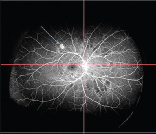
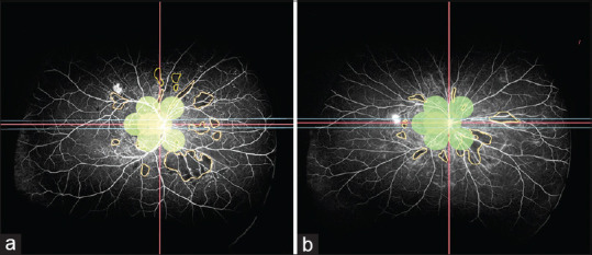
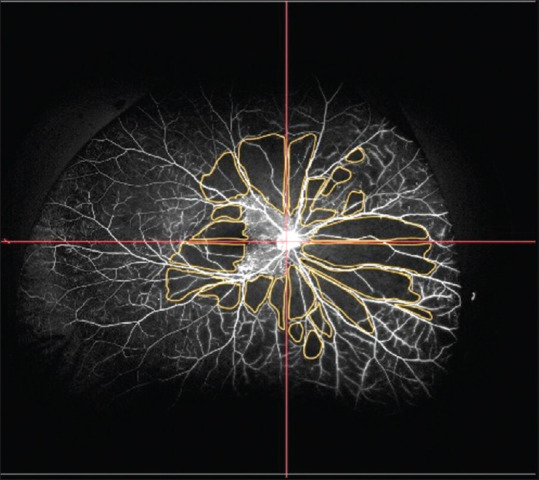
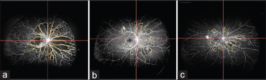
To analyze the topographic distribution of neovascularization (NV) and capillary nonperfusion (CNP) using ultra-wide field fluorescein angiography (UWFFA) in patients with proliferative diabetic retinopathy (PDR).

METHODS

This was a prospective, single-center, observational study in which all patients who presented between March 2019 and December 2020 and satisfied the inclusion criteria were recruited. In our study, patients with treatment-naïve PDR without any fibrovascular proliferation underwent UWFFA. The images were analyzed qualitatively for the topographic distribution of NV and the CNP area was quantified. The number of lesions picked by UWFFA was compared with 7 standard field (7SF) image using overlay of 7SF. The main outcome measure was characteristics of neovascularization, such as the number, location, and area of CNP, measured using UWFFA, which was considered with 95% confidence intervals (CI).

RESULTS

Two hundred and fifty-three eyes of 187 patients with a mean age of 56.03 ± 8 years were included. Mean neovascularization elsewhere (NVE) was 2.91 ± 3.43. Maximum NVEs were seen in the superotemporal (ST; 0.9 ± 1.13) quadrant, followed by the inferotemporal (IT; 0.7 ± 1.08), inferonasal (IN; 0.66 ± 1.02) and superonasal (SN; 0.66 ± 1.01) quadrants. Maximum CNP area was seen in the SN (13.75 ± 8.83 disc diameter square [DD]) quadrant, followed by the IN (13.48 ± 8.59 DD), IT (11.34 ± 8.37 DD), and ST (11.3 ± 8.34 DD) quadrants. Mean CNP area was maximum in patients with only neovascularization of disc (NVD; 64.99 ± 41.47 DD), followed by both NVD and NVE (61.37 ± 35.61 DD), and was minimum in patients with only NVE (36.44 ± 22.03 DD). Eighty-one (32%) eyes out of 253 had NVE and 189 (75%) out of 253 had CNP area outside 7SF (overlay) of Early Treatment Diabetic Retinopathy Study (ETDRS).

CONCLUSION

Diabetic NV lesions and CNP areas are distributed asymmetrically throughout the retina and are not restricted to the posterior pole. Compared to conventional 7SF imaging, UWFFA reveals significantly more retinal vascular pathology in patients with PDR.

摘要

目的

利用超广角荧光素血管造影(UWFFA)分析增生型糖尿病视网膜病变(PDR)患者的新生血管(NV)和无灌注毛细血管(CNP)的地形分布。

方法

这是一项前瞻性、单中心、观察性研究,招募了 2019 年 3 月至 2020 年 12 月期间出现并符合纳入标准的所有患者。在我们的研究中,对未经治疗的无任何纤维血管增生的 PDR 患者进行 UWFFA。使用 7 标准视野(7SF)图像叠加对图像进行定性分析,以评估 NV 和 CNP 区域的地形分布,并对 UWFFA 检测到的病变数量与 7SF 进行比较。主要观察指标是 NV 的特征,如 CNP 的数量、位置和面积,采用 UWFFA 进行测量,置信区间(CI)为 95%。

结果

共纳入 187 例患者的 253 只眼,平均年龄为 56.03±8 岁。平均其他部位的新生血管(NVE)为 2.91±3.43。最大的 NVEs 见于上颞(ST;0.9±1.13)象限,其次是下颞(IT;0.7±1.08)、下鼻(IN;0.66±1.02)和上鼻(SN;0.66±1.01)象限。最大的 CNP 面积见于 SN(13.75±8.83 视盘直径平方[DD])象限,其次是 IN(13.48±8.59 DD)、IT(11.34±8.37 DD)和 ST(11.3±8.34 DD)象限。仅存在视盘新生血管(NVD)的患者的平均 CNP 面积最大(64.99±41.47 DD),其次是同时存在 NVD 和 NVE(61.37±35.61 DD),仅存在 NVE 的患者最小(36.44±22.03 DD)。253 只眼中,81 只(32%)有 NVE,253 只眼中 189 只(75%)有 7SF 以外的 CNP 区域(ETDRS 早期糖尿病视网膜病变研究的叠加)。

结论

糖尿病性 NV 病变和 CNP 区域在整个视网膜呈不对称分布,并不局限于后极部。与传统的 7SF 成像相比,UWFFA 显示 PDR 患者的视网膜血管病变明显更多。

https://cdn.ncbi.nlm.nih.gov/pmc/blobs/71a3/10538809/58bff32c83e7/IJO-71-3080-g004.jpg
https://cdn.ncbi.nlm.nih.gov/pmc/blobs/71a3/10538809/dfafd063d102/IJO-71-3080-g001.jpg
https://cdn.ncbi.nlm.nih.gov/pmc/blobs/71a3/10538809/e967a71f8dc7/IJO-71-3080-g002.jpg
https://cdn.ncbi.nlm.nih.gov/pmc/blobs/71a3/10538809/3ed1f883f540/IJO-71-3080-g003.jpg
https://cdn.ncbi.nlm.nih.gov/pmc/blobs/71a3/10538809/58bff32c83e7/IJO-71-3080-g004.jpg

相似文献

1
Topographic distribution of retinal neovascularization in proliferative diabetic retinopathy using ultra-wide field angiography.应用超广角血管造影观察增生性糖尿病视网膜病变的视网膜新生血管的分布。
Indian J Ophthalmol. 2023 Aug;71(8):3080-3084. doi: 10.4103/IJO.IJO_3285_22.
2
Retinal Nonperfusion Characteristics on Ultra-Widefield Angiography in Eyes With Severe Nonproliferative Diabetic Retinopathy and Proliferative Diabetic Retinopathy.在严重非增生性糖尿病性视网膜病变和增生性糖尿病性视网膜病变的眼中,超广角血管造影的视网膜无灌注特征。
JAMA Ophthalmol. 2019 Jun 1;137(6):626-631. doi: 10.1001/jamaophthalmol.2019.0440.
3
Characteristics of Retinal Neovascularization in Proliferative Diabetic Retinopathy Imaged by Optical Coherence Tomography Angiography.光学相干断层扫描血管造影成像的增殖性糖尿病视网膜病变中视网膜新生血管的特征
Invest Ophthalmol Vis Sci. 2016 Nov 1;57(14):6247-6255. doi: 10.1167/iovs.16-20210.
4
WF SS-OCTA for detecting diabetic retinopathy and evaluating the effect of photocoagulation on posterior vitreous detachment.应用 WF SS-OCTA 检测糖尿病视网膜病变并评估光凝对玻璃体后脱离的疗效。
Front Endocrinol (Lausanne). 2022 Dec 2;13:1029066. doi: 10.3389/fendo.2022.1029066. eCollection 2022.
5
Distribution of Nonperfusion and Neovascularization on Ultrawide-Field Fluorescein Angiography in Proliferative Diabetic Retinopathy (RECOVERY Study): Report 1.在增生型糖尿病视网膜病变(RECOVERY 研究)的超广角荧光素血管造影中,无灌注和新生血管的分布:报告 1。
Am J Ophthalmol. 2019 Oct;206:154-160. doi: 10.1016/j.ajo.2019.04.023. Epub 2019 May 10.
6
Distribution of Diabetic Neovascularization on Ultra-Widefield Fluorescein Angiography and on Simulated Widefield OCT Angiography.超广角荧光素血管造影和模拟广角 OCT 血管造影上糖尿病新生血管的分布。
Am J Ophthalmol. 2019 Nov;207:110-120. doi: 10.1016/j.ajo.2019.05.031. Epub 2019 Jun 10.
7
Wide-field optical coherence tomography angiography for the detection of proliferative diabetic retinopathy.宽视野光学相干断层扫描血管造影在增殖性糖尿病视网膜病变中的应用。
Graefes Arch Clin Exp Ophthalmol. 2020 Sep;258(9):1901-1909. doi: 10.1007/s00417-020-04773-x. Epub 2020 May 30.
8
Comparison between wide-angle OCT angiography and ultra-wide field fluorescein angiography for detecting non-perfusion areas and retinal neovascularization in eyes with diabetic retinopathy.广角光学相干断层扫描血管造影术与超广角荧光素血管造影术在检测糖尿病视网膜病变患者眼部无灌注区和视网膜新生血管方面的比较。
Graefes Arch Clin Exp Ophthalmol. 2018 Jul;256(7):1275-1280. doi: 10.1007/s00417-018-3992-y. Epub 2018 Apr 30.
9
Feasibility and Clinical Utility of Wide-Field Optical Coherence Tomography Angiography Compared to Ultrawide-Field Fluorescein Angiography in Patients with Diabetic Retinopathy.宽视野光学相干断层扫描血管造影与超广角荧光素血管造影在糖尿病视网膜病变患者中的可行性和临床应用比较。
Klin Monbl Augenheilkd. 2023 Apr;240(4):490-495. doi: 10.1055/a-2031-2657. Epub 2023 Apr 25.
10
Longitudinal Wide-Field Swept-Source OCT Angiography of Neovascularization in Proliferative Diabetic Retinopathy after Panretinal Photocoagulation.全视网膜光凝术后增殖性糖尿病视网膜病变新生血管形成的纵向广角扫频源光学相干断层扫描血管造影
Ophthalmol Retina. 2019 Apr;3(4):350-361. doi: 10.1016/j.oret.2018.11.008. Epub 2018 Nov 24.

引用本文的文献

1
Author's Response: Ultrawide field angiography in proliferative diabetic retinopathy.作者回复:增殖性糖尿病视网膜病变中的超广角血管造影
Indian J Ophthalmol. 2024 Nov 1;72(11):1676-1677. doi: 10.4103/IJO.IJO_1058_24. Epub 2024 Oct 26.
2
Differential distribution of fibrovascular proliferative membranes in 25-gauge vitrectomy for proliferative diabetic retinopathy.25G玻璃体切割术治疗增殖性糖尿病视网膜病变时纤维血管增殖膜的差异分布
Int J Ophthalmol. 2024 Aug 18;17(8):1462-1468. doi: 10.18240/ijo.2024.08.11. eCollection 2024.
3
Regulation role of miR-204 on SIRT1/VEGF in metabolic memory induced by high glucose in human retinal pigment epithelial cells.

本文引用的文献

1
Distribution of Diabetic Neovascularization on Ultra-Widefield Fluorescein Angiography and on Simulated Widefield OCT Angiography.超广角荧光素血管造影和模拟广角 OCT 血管造影上糖尿病新生血管的分布。
Am J Ophthalmol. 2019 Nov;207:110-120. doi: 10.1016/j.ajo.2019.05.031. Epub 2019 Jun 10.
2
Distribution of Nonperfusion and Neovascularization on Ultrawide-Field Fluorescein Angiography in Proliferative Diabetic Retinopathy (RECOVERY Study): Report 1.在增生型糖尿病视网膜病变(RECOVERY 研究)的超广角荧光素血管造影中,无灌注和新生血管的分布:报告 1。
Am J Ophthalmol. 2019 Oct;206:154-160. doi: 10.1016/j.ajo.2019.04.023. Epub 2019 May 10.
3
miR-204对人视网膜色素上皮细胞中高糖诱导的代谢记忆中SIRT1/VEGF的调控作用
Int J Ophthalmol. 2024 Jul 18;17(7):1232-1237. doi: 10.18240/ijo.2024.07.06. eCollection 2024.
4
Ultrawide field angiography in proliferative diabetic retinopathy.增殖性糖尿病视网膜病变的超广角血管造影
Indian J Ophthalmol. 2024 May 1;72(5):754. doi: 10.4103/IJO.IJO_2416_23. Epub 2024 Apr 22.
Extended targeted retinal photocoagulation versus conventional pan-retinal photocoagulation for proliferative diabetic retinopathy in a randomized clinical trial.
一项随机临床试验中,对比扩展靶向视网膜光凝与传统全视网膜光凝治疗增殖性糖尿病视网膜病变的疗效
Int Ophthalmol. 2018 Feb;38(1):313-321. doi: 10.1007/s10792-017-0469-7. Epub 2017 Feb 6.
4
Update on wide- and ultra-widefield retinal imaging.广角和超广角视网膜成像的最新进展。
Indian J Ophthalmol. 2015 Jul;63(7):575-81. doi: 10.4103/0301-4738.167122.
5
New vessels detected on wide-field imaging compared to two-field and seven-field imaging: implications for diabetic retinopathy screening image analysis.与双视野和七视野成像相比,在广角成像中检测到的新血管:对糖尿病视网膜病变筛查图像分析的影响。
Br J Ophthalmol. 2015 Dec;99(12):1606-9. doi: 10.1136/bjophthalmol-2015-306719. Epub 2015 Aug 13.
6
Optomap ultrawide field imaging identifies additional retinal abnormalities in patients with diabetic retinopathy.Optomap超广角成像可识别糖尿病视网膜病变患者的其他视网膜异常。
Clin Ophthalmol. 2015 Mar 24;9:527-31. doi: 10.2147/OPTH.S79448. eCollection 2015.
7
Peripheral lesions identified by mydriatic ultrawide field imaging: distribution and potential impact on diabetic retinopathy severity.散瞳超广角成像识别的周边病变:分布及对糖尿病视网膜病变严重程度的潜在影响。
Ophthalmology. 2013 Dec;120(12):2587-2595. doi: 10.1016/j.ophtha.2013.05.004. Epub 2013 Jun 15.
8
Topographical distribution of retinal and optic disc neovascularization in early stages of proliferative diabetic retinopathy.增生型糖尿病视网膜病变早期视网膜和视盘新生血管的分布。
Invest Ophthalmol Vis Sci. 2012 Dec 17;53(13):8246-52. doi: 10.1167/iovs.12-10918.
9
Ultra-wide-field angiography improves the detection and classification of diabetic retinopathy.超广角血管造影提高糖尿病视网膜病变的检出率和分类准确率。
Retina. 2012 Apr;32(4):785-91. doi: 10.1097/IAE.0b013e3182278b64.
10
The topographic distribution of the first sites of diabetic retinal neovascularization.糖尿病视网膜新生血管形成首发部位的地形图分布
Am J Ophthalmol. 1998 May;125(5):704-6. doi: 10.1016/s0002-9394(98)00013-0.