Liu Yufeng, Jiang Shengdian, Li Yingxin, Zhao Sujun, Yun Zhixi, Zhao Zuo-Han, Zhang Lingli, Wang Gaoyu, Chen Xin, Manubens-Gil Linus, Hang Yuning, Garcia-Forn Marta, Wang Wei, Rubeis Silvia De, Wu Zhuhao, Osten Pavel, Gong Hui, Hawrylycz Michael, Mitra Partha, Dong Hongwei, Luo Qingming, Ascoli Giorgio A, Zeng Hongkui, Liu Lijuan, Peng Hanchuan
SEU-ALLEN Joint Center, Institute for Brain and Intelligence, Southeast University, Nanjing, China.
Seaver Autism Center for Research and Treatment, Icahn School of Medicine at Mount Sinai, New York, NY, USA.
Res Sq. 2023 Jul 25:rs.3.rs-3146034. doi: 10.21203/rs.3.rs-3146034/v1.
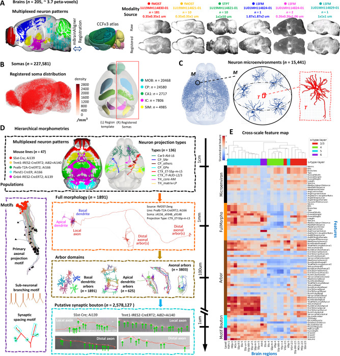
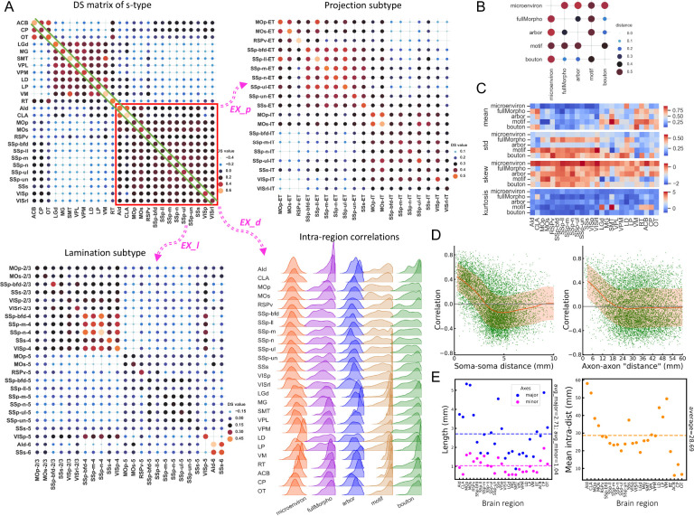
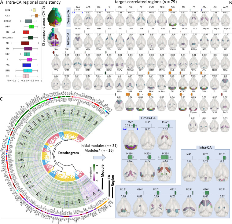
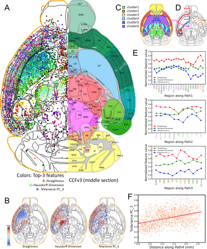
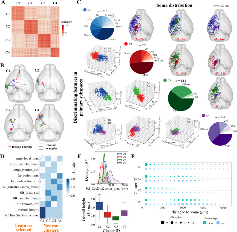
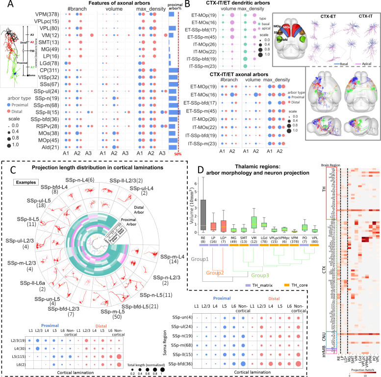
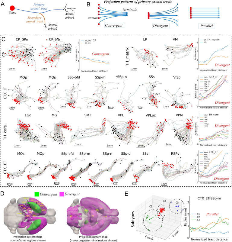
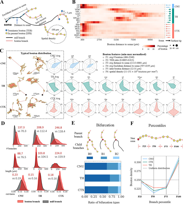
We conducted a large-scale study of whole-brain morphometry, analyzing 3.7 peta-voxels of mouse brain images at the single-cell resolution, producing one of the largest multi-morphometry databases of mammalian brains to date. We spatially registered 205 mouse brains and associated data from six Brain Initiative Cell Census Network (BICCN) data sources covering three major imaging modalities from five collaborative projects to the Allen Common Coordinate Framework (CCF) atlas, annotated 3D locations of cell bodies of 227,581 neurons, modeled 15,441 dendritic microenvironments, characterized the full morphology of 1,891 neurons along with their axonal motifs, and detected 2.58 million putative synaptic boutons. Our analysis covers six levels of information related to neuronal populations, dendritic microenvironments, single-cell full morphology, sub-neuronal dendritic and axonal arborization, axonal boutons, and structural motifs, along with a quantitative characterization of the diversity and stereotypy of patterns at each level. We identified 16 modules consisting of highly intercorrelated brain regions in 13 functional brain areas corresponding to 314 anatomical regions in CCF. Our analysis revealed the dendritic microenvironment as a powerful method for delineating brain regions of cell types and potential subtypes. We also found that full neuronal morphologies can be categorized into four distinct classes based on spatially tuned morphological features, with substantial cross-areal diversity in apical dendrites, basal dendrites, and axonal arbors, along with quantified stereotypy within cortical, thalamic and striatal regions. The lamination of somas was found to be more effective in differentiating neuron arbors within the cortex. Further analysis of diverging and converging projections of individual neurons in 25 regions throughout the brain reveals branching preferences in the brain-wide and local distributions of axonal boutons. Overall, our study provides a comprehensive description of key anatomical structures of neurons and their types, covering a wide range of scales and features, and contributes to our understanding of neuronal diversity and its function in the mammalian brain.
我们进行了一项全脑形态测量的大规模研究,以单细胞分辨率分析了3.7拍字节的小鼠脑图像,生成了迄今为止最大的哺乳动物脑多形态测量数据库之一。我们将205个小鼠脑以及来自六个脑计划细胞普查网络(BICCN)数据源的相关数据进行空间配准,这些数据涵盖了来自五个合作项目的三种主要成像模式,并将其配准到艾伦通用坐标框架(CCF)图谱上,标注了227581个神经元细胞体的三维位置,对15441个树突微环境进行建模,描绘了1891个神经元及其轴突模式的完整形态,并检测到258万个假定的突触小体。我们的分析涵盖了与神经元群体、树突微环境、单细胞完整形态、亚神经元树突和轴突分支、轴突小体以及结构基序相关的六个信息层面,同时还对每个层面模式的多样性和刻板性进行了定量表征。我们在与CCF中314个解剖区域相对应的13个功能脑区中识别出由高度相互关联的脑区组成的16个模块。我们的分析表明,树突微环境是描绘细胞类型和潜在亚型脑区的有力方法。我们还发现,基于空间调整的形态特征,完整的神经元形态可分为四个不同类别,在顶树突、基底树突和轴突分支中存在大量跨区域多样性,同时在皮质、丘脑和纹状体区域内存在量化的刻板性。发现体细胞的分层在区分皮质内的神经元分支方面更有效。对全脑25个区域中单个神经元的发散和汇聚投射的进一步分析揭示了轴突小体在全脑和局部分布中的分支偏好。总体而言,我们的研究全面描述了神经元及其类型的关键解剖结构,涵盖了广泛的尺度和特征,并有助于我们理解哺乳动物脑中神经元的多样性及其功能。