Department of Ophthalmology & Visual Science, College of Medicine, St. Vincent's Hospital, The Catholic University of Korea, Seoul, Republic of Korea.
Department of Ophthalmology & Visual Science, College of Medicine, Seoul St. Mary's Hospital, The Catholic University of Korea, Seoul, Republic of Korea.
Sci Rep. 2023 Aug 8;13(1):12879. doi: 10.1038/s41598-023-40025-8.
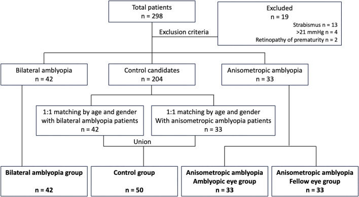
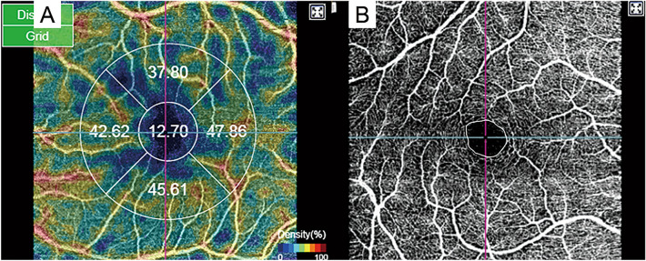
We analyzed whether macular superficial vascular density (SVD) and foveal vascular zone (FAZ) on optical coherence tomography angiography (OCTA) can distinguish between bilateral ametropic and anisometropic amblyopia. We included 42, 33, and 50 eyes in the bilateral ametropic amblyopia, anisometropic amblyopia, and normal control groups, respectively. Using macular swept-source optical coherence tomography angiography, we measured and analyzed the superficial FAZ areas and five sectoral macular SVDs after magnification correction. The anisometropic amblyopic eye group showed significantly increased foveal SVDs (p < 0.001) and significantly decreased superficial FAZ areas (p < 0.001), compared with the remaining groups. Additionally, the bilateral ametropic amblyopia group had significantly decreased nasal SVDs. SVDs and superficial FAZ areas differed among hyperopic amblyopia subtypes. These findings may reflect vascular distribution differences and macular changes in hyperopic amblyopia subtypes compared with normal eyes.
我们分析了光学相干断层扫描血管造影(OCTA)上的黄斑浅层血管密度(SVD)和中心凹血管区(FAZ)是否可以区分双眼屈光不正性和非屈光不正性弱视。我们分别纳入了双眼屈光不正性弱视、非屈光不正性弱视和正常对照组中的 42、33 和 50 只眼。使用黄斑扫频源 OCTA,我们在放大矫正后测量和分析了浅层 FAZ 区域和五个扇形黄斑 SVD。与其他组相比,非屈光不正性弱视眼组的中心凹 SVD 显著增加(p<0.001),浅层 FAZ 区域显著减小(p<0.001)。此外,双眼屈光不正性弱视组的鼻侧 SVD 显著降低。不同类型远视性弱视的 SVD 和浅层 FAZ 区域存在差异。这些发现可能反映了远视性弱视亚型与正常眼相比在血管分布和黄斑变化方面的差异。