Gerken Annika, Walluscheck Sina, Kohlmann Peter, Galinovic Ivana, Villringer Kersten, Fiebach Jochen B, Klein Jan, Heldmann Stefan
Fraunhofer Institute for Digital Medicine MEVIS, Bremen, Germany.
Fraunhofer Institute for Digital Medicine MEVIS, Lübeck, Germany.
Front Neuroimaging. 2023 Aug 4;2:1228255. doi: 10.3389/fnimg.2023.1228255. eCollection 2023.
The automatic segmentation of brain parenchyma and cerebrospinal fluid-filled spaces such as the ventricular system is the first step for quantitative and qualitative analysis of brain CT data. For clinical practice and especially for diagnostics, it is crucial that such a method is robust to anatomical variability and pathological changes such as (hemorrhagic or neoplastic) lesions and chronic defects. This study investigates the increase in overall robustness of a deep learning algorithm that is gained by adding hemorrhage training data to an otherwise normal training cohort.
A 2D U-Net is trained on subjects with normal appearing brain anatomy. In a second experiment the training data includes additional subjects with brain hemorrhage on image data of the RSNA Brain CT Hemorrhage Challenge with custom reference segmentations. The resulting networks are evaluated on normal and hemorrhage test casesseparately, and on an independent test set of patients with brain tumors of the publicly available GLIS-RT dataset.
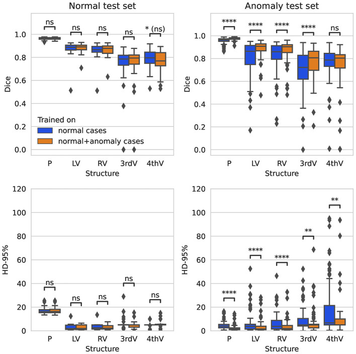
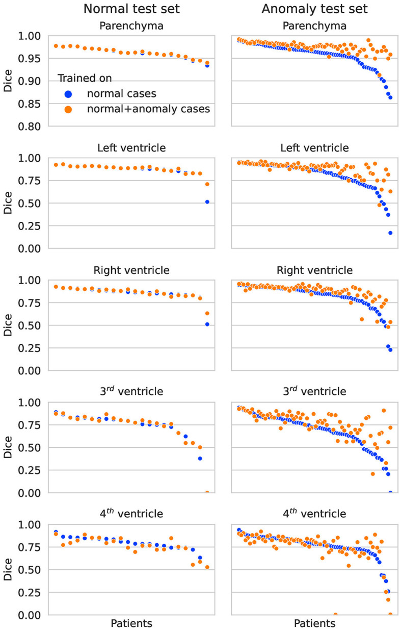
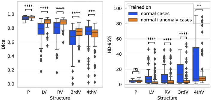
Adding data with hemorrhage to the training set significantly improves the segmentation performance over an algorithm trained exclusively on normally appearing data, not only in the hemorrhage test set but also in the tumor test set. The performance on normally appearing data is stable. Overall, the improved algorithm achieves median Dice scores of 0.98 (parenchyma), 0.91 (left ventricle), 0.90 (right ventricle), 0.81 (third ventricle), and 0.80 (fourth ventricle) on the hemorrhage test set. On the tumor test set, the median Dice scores are 0.96 (parenchyma), 0.90 (left ventricle), 0.90 (right ventricle), 0.75 (third ventricle), and 0.73 (fourth ventricle).
Training on an extended data set that includes pathologies is crucial and significantly increases the overall robustness of a segmentation algorithm for brain parenchyma and ventricular system in CT data, also for anomalies completely unseen during training. Extension of the training set to include other diseases may further improve the generalizability of the algorithm.
脑实质和脑脊液填充空间(如脑室系统)的自动分割是对脑部CT数据进行定量和定性分析的第一步。对于临床实践,尤其是诊断而言,这样一种方法对于解剖变异和病理变化(如(出血性或肿瘤性)病变和慢性缺损)具有鲁棒性至关重要。本研究调查了通过向原本正常的训练队列中添加出血训练数据而获得的深度学习算法整体鲁棒性的提升。
在具有正常脑解剖结构的受试者上训练二维U-Net。在第二个实验中,训练数据包括来自RSNA脑部CT出血挑战赛图像数据且带有定制参考分割的额外脑出血受试者。分别在正常和出血测试病例以及公开可用的GLIS-RT数据集中脑肿瘤患者的独立测试集上评估所得网络。
在训练集中添加出血数据相较于仅在正常外观数据上训练的算法,显著提高了分割性能,不仅在出血测试集中如此,在肿瘤测试集中也是如此。在正常外观数据上的性能稳定。总体而言,改进后的算法在出血测试集上脑实质、左心室、右心室、第三脑室和第四脑室的中位Dice分数分别为0.98、0.91、0.90、0.81和0.80。在肿瘤测试集上,中位Dice分数分别为0.96、0.90、0.90、0.75和0.73。
在包含病理学数据的扩展数据集上进行训练至关重要,并且显著提高了CT数据中脑实质和脑室系统分割算法的整体鲁棒性,对于训练期间完全未见过的异常情况也是如此。将训练集扩展以纳入其他疾病可能会进一步提高该算法的通用性。