Department of Radiological, Oncological and Pathological Sciences, Umberto I Hospital, Sapienza University of Rome, Rome, Italy.
National Research Council (CNR),, Institute for Complex Systems (ISC) c/o Physics Department Sapienza University of Rome, Rome, Italy.
Eur Radiol Exp. 2023 Aug 10;7(1):41. doi: 10.1186/s41747-023-00358-5.
Fetal magnetic resonance imaging (fetal MRI) is usually performed as a second-level examination following routine ultrasound examination, generally exploiting morphological and diffusion MRI sequences. The objective of this review is to describe the novelties and new applications of fetal MRI, focusing on three main aspects: the new sequences with their applications, the transition from 1.5-T to 3-T magnetic field, and the new applications of artificial intelligence software. This review was carried out by consulting the MEDLINE references (PubMed) and including only peer-reviewed articles written in English. Among the most important novelties in fetal MRI, we find the intravoxel incoherent motion model which allow to discriminate the diffusion from the perfusion component in fetal and placenta tissues. The transition from 1.5-T to 3-T magnetic field allowed for higher quality images, thanks to the higher signal-to-noise ratio with a trade-off of more frequent artifacts. The application of motion-correction software makes it possible to overcome movement artifacts by obtaining higher quality images and to generate three-dimensional images useful in preoperative planning.Relevance statementThis review shows the latest developments offered by fetal MRI focusing on new sequences, transition from 1.5-T to 3-T magnetic field and the emerging role of AI software that are paving the way for new diagnostic strategies.Key points• Fetal magnetic resonance imaging (MRI) is a second-line imaging after ultrasound.• Diffusion-weighted imaging and intravoxel incoherent motion sequences provide quantitative biomarkers on fetal microstructure and perfusion.• 3-T MRI improves the detection of cerebral malformations.• 3-T MRI is useful for both body and nervous system indications.• Automatic MRI motion tracking overcomes fetal movement artifacts and improve fetal imaging.
胎儿磁共振成像(fetal MRI)通常作为常规超声检查后的二线检查,通常利用形态学和扩散 MRI 序列进行。本综述的目的是描述胎儿 MRI 的新进展和新应用,重点关注三个主要方面:新序列及其应用、从 1.5-T 到 3-T 磁场的转变,以及人工智能软件的新应用。本综述通过查阅 MEDLINE 参考文献(PubMed)并仅纳入以英文撰写的同行评审文章来进行。在胎儿 MRI 的最重要的新进展中,我们发现了体素内不相干运动模型,该模型可区分胎儿和胎盘组织中的扩散和灌注成分。从 1.5-T 到 3-T 磁场的转变允许获得更高质量的图像,这要归功于更高的信噪比,但代价是更频繁的伪影。运动校正软件的应用使得通过获得更高质量的图像来克服运动伪影并生成在术前规划中有用的三维图像成为可能。
相关性声明
本综述展示了胎儿 MRI 的最新发展,重点关注新序列、从 1.5-T 到 3-T 磁场的转变以及人工智能软件的新兴作用,这些都为新的诊断策略铺平了道路。
要点
• 胎儿磁共振成像(MRI)是超声检查后的二线成像方法。
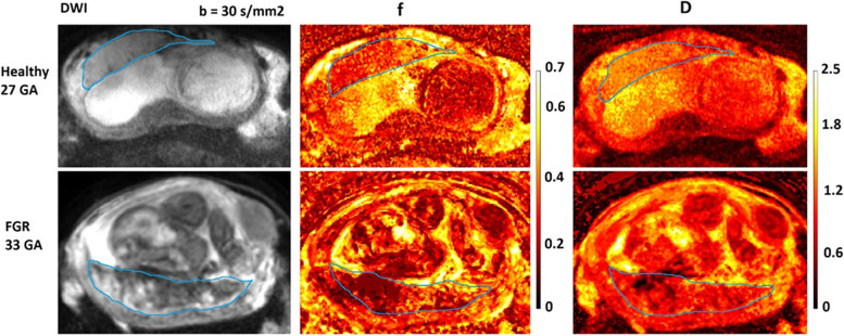
• 扩散加权成像和体素内不相干运动序列为胎儿微观结构和灌注提供定量生物标志物。
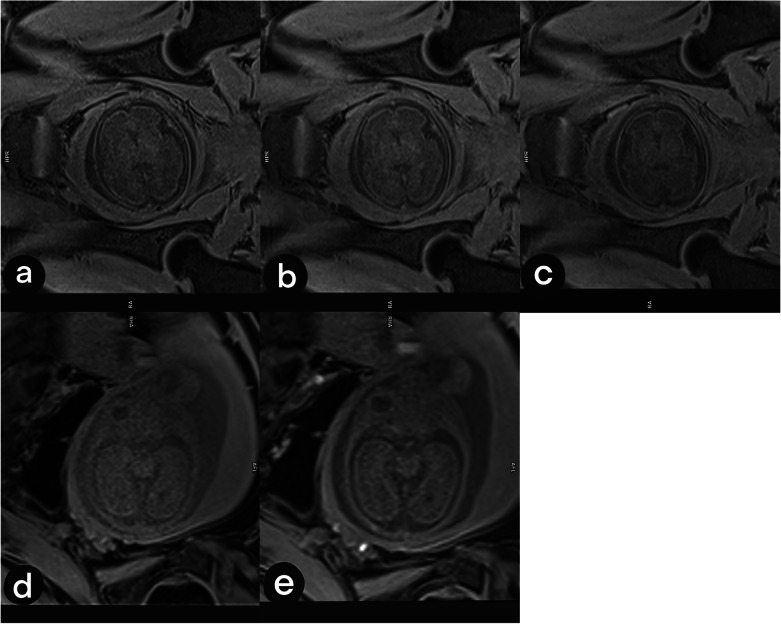
• 3-T MRI 提高了对脑畸形的检测能力。
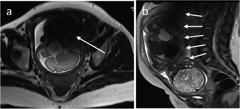
• 3-T MRI 对身体和神经系统适应证均有用。
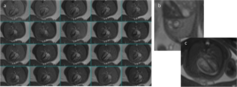
• 自动 MRI 运动跟踪克服了胎儿运动伪影,改善了胎儿成像。