Cundari Giulia, Galea Nicola, Di Mascio Daniele, Gennarini Marco, Ventriglia Flavia, Curti Federica, Dodaro Martina, Rizzo Giuseppe, Catalano Carlo, Giancotti Antonella, Manganaro Lucia
Department of Radiological, Oncological and Pathological Sciences, Sapienza University of Rome, Policlinico Umberto I, Viale Regina Elena 324, 00161 Rome, Italy.
Department of Maternal and Child Health and Urological Sciences, Sapienza University of Rome, Policlinico Umberto I, Viale Regina Elena 324, 00161 Rome, Italy.
J Clin Med. 2024 Aug 6;13(16):4598. doi: 10.3390/jcm13164598.
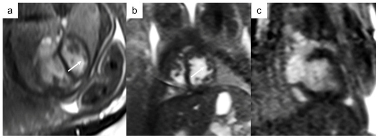
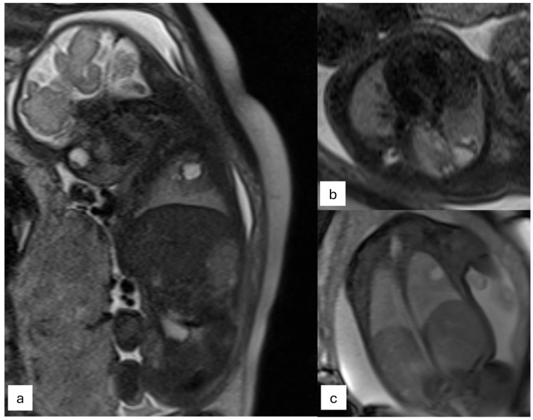
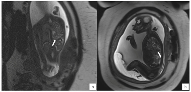
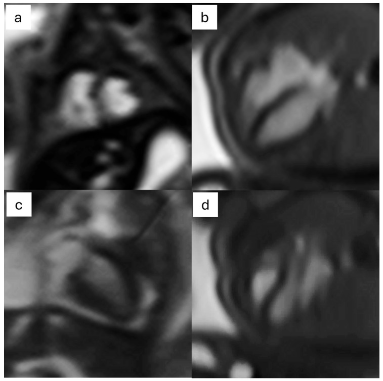
Fetal magnetic resonance imaging (fMRI) represents a second-line imaging modality that provides multiparametric and multiplanar views that are crucial for confirming diagnoses, detecting associated pathologies, and resolving inconclusive ultrasound findings. The introduction of high-field magnets and new imaging sequences has expanded MRI's role in pregnancy management. Recent innovations in ECG-gating techniques have revolutionized the prenatal evaluation of congenital heart disease by synchronizing imaging with the fetal heartbeat, thus addressing traditional challenges in cardiac imaging. Fetal cardiac MRI (fCMR) is particularly valuable for assessing congenital heart diseases, especially when ultrasound is limited by poor imaging conditions. fCMR allows for detailed anatomical and functional evaluation of the heart and great vessels and is also useful for diagnosing additional anomalies and analyzing blood flow patterns, which can aid in understanding abnormal fetal brain growth and placental perfusion. This review emphasizes fMRI's potential in evaluating cardiac and thoracic structures, including various gating techniques like metric optimized gating, self-gating, and Doppler ultrasound gating. The review also covers the use of static and cine images for structural and functional assessments and discusses advanced techniques like 4D-flow MRI and T1 or T2 mapping for comprehensive flow quantification and tissue characterization.
胎儿磁共振成像(fMRI)是一种二线成像方式,它能提供多参数和多平面视图,这对于确诊、检测相关病变以及解决超声检查结果不明确的情况至关重要。高场强磁体和新成像序列的引入扩大了MRI在孕期管理中的作用。心电图门控技术的最新创新通过使成像与胎儿心跳同步,彻底改变了先天性心脏病的产前评估,从而解决了心脏成像中的传统难题。胎儿心脏磁共振成像(fCMR)在评估先天性心脏病方面特别有价值,尤其是当超声受成像条件不佳限制时。fCMR能够对心脏和大血管进行详细的解剖和功能评估,也有助于诊断其他异常情况并分析血流模式,这有助于理解胎儿脑生长异常和胎盘灌注情况。本综述强调了fMRI在评估心脏和胸部结构方面的潜力,包括各种门控技术,如度量优化门控、自门控和多普勒超声门控。该综述还涵盖了使用静态和电影图像进行结构和功能评估,并讨论了诸如4D流MRI和T1或T2映射等先进技术,用于全面的血流定量和组织特征分析。