Department of Pathology, Peking Union Medical College Hospital, Chinese Academy of Medical Sciences and Peking Union Medical College, Beijing 100730, China.
State Key Laboratory of Bioactive Substance and Function of Natural Medicines, Institute of Materia Medica, Chinese Academy of Medical Sciences and Peking Union Medical College, Beijing 100050, China.
Molecules. 2023 Jul 31;28(15):5791. doi: 10.3390/molecules28155791.
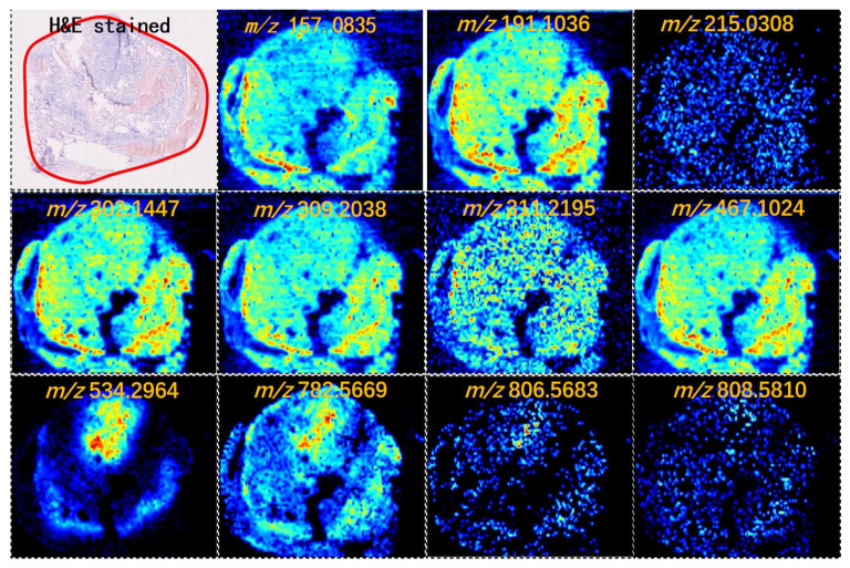
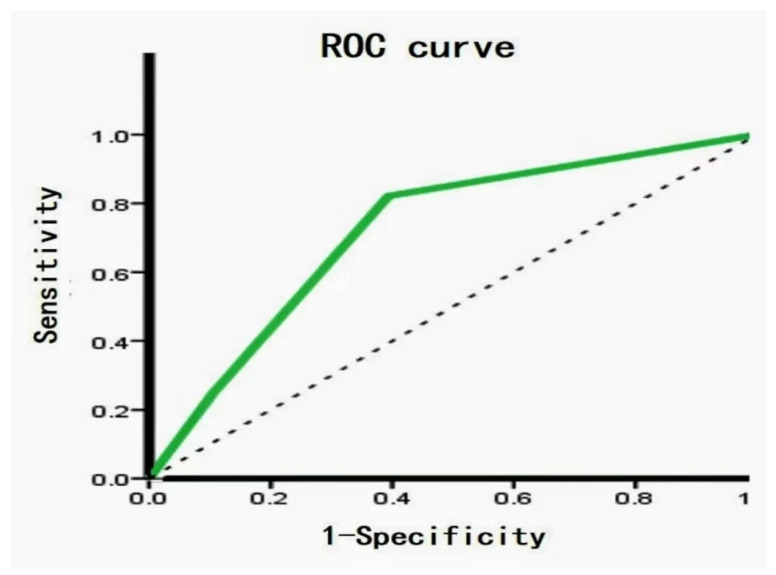
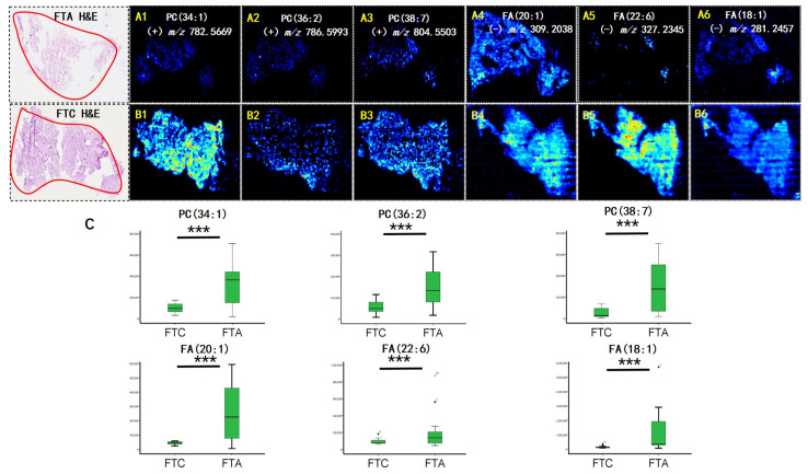
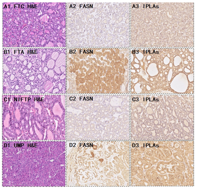
"Gray zone" thyroid follicular tumors are difficult to diagnose, especially when distinguishing between benign follicular thyroid adenoma (FTA) and malignant carcinoma (FTC). Thus, proper classification of thyroid follicular diseases may improve clinical prognosis. In this study, the diagnostic performance of metabolite enzymes was evaluated using imaging mass spectrometry to distinguish FTA from FTC and determine the association between metabolite enzyme expression with thyroid follicular borderline tumor diagnosis. Air flow-assisted desorption electrospray ionization mass spectrometry imaging (AFAIDESI-MSI) was used to build a classification model for thyroid follicular tumor characteristics among 24 samples. We analyzed metabolic enzyme marker expression in an independent validation set of 133 cases and further evaluated the potential biological behavior of 19 thyroid borderline lesions. Phospholipids and fatty acids (FAs) were more abundant in FTA than FTC ( < 0.001). The metabolic enzyme panel, which included FA synthase and Ca-independent PLA2, was further validated in follicular thyroid tumors. The marker combination showed optimal performance in the validation group (area under the ROC, sensitivity, and specificity: 73.6%, 82.1%, and 60.6%, respectively). The findings indicate that AFAIDESI-MSI, in combination with low metabolic enzyme expression, could play a role in the diagnosis of thyroid follicular borderline tumors for strict follow-up.
“灰色区域”甲状腺滤泡性肿瘤难以诊断,特别是在区分良性滤泡状甲状腺腺瘤(FTA)和恶性癌(FTC)时。因此,对甲状腺滤泡性疾病进行适当分类可能会改善临床预后。在这项研究中,使用成像质谱法评估代谢酶的诊断性能,以区分 FTA 和 FTC,并确定代谢酶表达与甲状腺滤泡交界性肿瘤诊断之间的关联。采用气流辅助解吸电喷雾电离质谱成像(AFAIDESI-MSI)技术,对 24 例样本的甲状腺滤泡肿瘤特征构建分类模型。我们分析了 133 例独立验证集中代谢酶标志物的表达,并进一步评估了 19 例甲状腺交界性病变的潜在生物学行为。FTA 中的磷脂和脂肪酸(FA)比 FTC 更为丰富(<0.001)。在甲状腺滤泡肿瘤中进一步验证了包括脂肪酸合酶和钙非依赖性 PLA2 在内的代谢酶谱。该标志物组合在验证组中表现出最佳性能(ROC 曲线下面积、敏感性和特异性分别为 73.6%、82.1%和 60.6%)。这些发现表明,AFAIDESI-MSI 结合低代谢酶表达,可能在严格随访的甲状腺滤泡交界性肿瘤的诊断中发挥作用。