• 文献检索
  • 文档翻译
  • 深度研究
  • 学术资讯
  • Suppr Zotero 插件Zotero 插件
  • 邀请有礼
  • 套餐&价格
  • 历史记录
应用&插件
Suppr Zotero 插件Zotero 插件浏览器插件Mac 客户端Windows 客户端微信小程序
定价
高级版会员购买积分包购买API积分包
服务
文献检索文档翻译深度研究API 文档MCP 服务
关于我们
关于 Suppr公司介绍联系我们用户协议隐私条款
关注我们

Suppr 超能文献

核心技术专利:CN118964589B侵权必究
粤ICP备2023148730 号-1Suppr @ 2026

文献检索

告别复杂PubMed语法,用中文像聊天一样搜索,搜遍4000万医学文献。AI智能推荐,让科研检索更轻松。

立即免费搜索

文件翻译

保留排版,准确专业,支持PDF/Word/PPT等文件格式,支持 12+语言互译。

免费翻译文档

深度研究

AI帮你快速写综述,25分钟生成高质量综述,智能提取关键信息,辅助科研写作。

立即免费体验

空间转录组学揭示了杜氏肌营养不良症小鼠模型中组织病理学变化的标志物。

Spatial transcriptomics reveal markers of histopathological changes in Duchenne muscular dystrophy mouse models.

机构信息

Department of Human Genetics, Leiden University Medical Center, Leiden, The Netherlands.

Department of Radiology, Leiden University Medical Center, Leiden, The Netherlands.

出版信息

Nat Commun. 2023 Aug 15;14(1):4909. doi: 10.1038/s41467-023-40555-9.

DOI:10.1038/s41467-023-40555-9
PMID:37582915
原文链接:https://pmc.ncbi.nlm.nih.gov/articles/PMC10427630/
Abstract

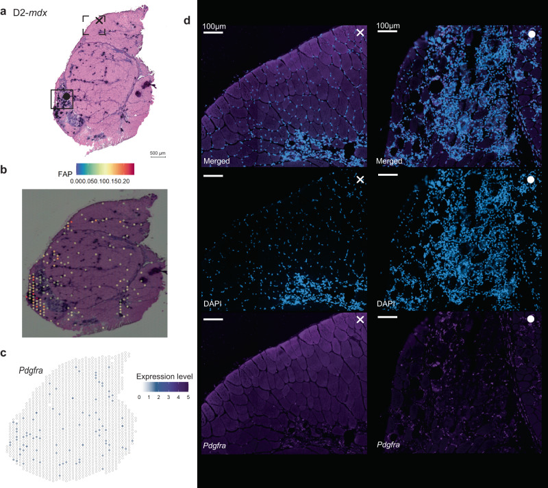
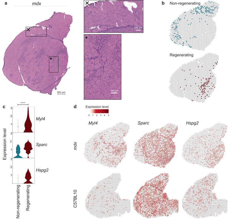
Duchenne muscular dystrophy is caused by mutations in the DMD gene, leading to lack of dystrophin. Chronic muscle damage eventually leads to histological alterations in skeletal muscles. The identification of genes and cell types driving tissue remodeling is a key step to developing effective therapies. Here we use spatial transcriptomics in two Duchenne muscular dystrophy mouse models differing in disease severity to identify gene expression signatures underlying skeletal muscle pathology and to directly link gene expression to muscle histology. We perform deconvolution analysis to identify cell types contributing to histological alterations. We show increased expression of specific genes in areas of muscle regeneration (Myl4, Sparc, Hspg2), fibrosis (Vim, Fn1, Thbs4) and calcification (Bgn, Ctsk, Spp1). These findings are confirmed by smFISH. Finally, we use differentiation dynamic analysis in the D2-mdx muscle to identify muscle fibers in the present state that are predicted to become affected in the future state.

摘要

杜氏肌营养不良症是由 DMD 基因突变引起的,导致肌营养不良蛋白缺失。慢性肌肉损伤最终导致骨骼肌的组织学改变。确定驱动组织重塑的基因和细胞类型是开发有效治疗方法的关键步骤。在这里,我们使用空间转录组学在两种疾病严重程度不同的杜氏肌营养不良症小鼠模型中进行研究,以确定骨骼肌肉病理学的基因表达特征,并将基因表达直接与肌肉组织学联系起来。我们进行去卷积分析以确定导致组织学改变的细胞类型。我们发现肌肉再生区域(Myl4、Sparc、Hspg2)、纤维化(Vim、Fn1、Thbs4)和钙化(Bgn、Ctsk、Spp1)中特定基因的表达增加。这些发现通过 smFISH 得到了证实。最后,我们在 D2-mdx 肌肉中使用分化动态分析来鉴定目前状态下的肌肉纤维,这些纤维预计在未来状态下会受到影响。

https://cdn.ncbi.nlm.nih.gov/pmc/blobs/aa4b/10427630/0d57bd216387/41467_2023_40555_Fig8_HTML.jpg
https://cdn.ncbi.nlm.nih.gov/pmc/blobs/aa4b/10427630/7e6bfcd9f646/41467_2023_40555_Fig1_HTML.jpg
https://cdn.ncbi.nlm.nih.gov/pmc/blobs/aa4b/10427630/bdabfdbeebac/41467_2023_40555_Fig2_HTML.jpg
https://cdn.ncbi.nlm.nih.gov/pmc/blobs/aa4b/10427630/b575b647c600/41467_2023_40555_Fig3_HTML.jpg
https://cdn.ncbi.nlm.nih.gov/pmc/blobs/aa4b/10427630/c7dfc090e9cd/41467_2023_40555_Fig4_HTML.jpg
https://cdn.ncbi.nlm.nih.gov/pmc/blobs/aa4b/10427630/6f2854b4e25a/41467_2023_40555_Fig5_HTML.jpg
https://cdn.ncbi.nlm.nih.gov/pmc/blobs/aa4b/10427630/168729c90a20/41467_2023_40555_Fig6_HTML.jpg
https://cdn.ncbi.nlm.nih.gov/pmc/blobs/aa4b/10427630/646f58de2d34/41467_2023_40555_Fig7_HTML.jpg
https://cdn.ncbi.nlm.nih.gov/pmc/blobs/aa4b/10427630/0d57bd216387/41467_2023_40555_Fig8_HTML.jpg

相似文献

1
Spatial transcriptomics reveal markers of histopathological changes in Duchenne muscular dystrophy mouse models.空间转录组学揭示了杜氏肌营养不良症小鼠模型中组织病理学变化的标志物。
Nat Commun. 2023 Aug 15;14(1):4909. doi: 10.1038/s41467-023-40555-9.
2
Long-Term Protective Effect of Human Dystrophin Expressing Chimeric (DEC) Cell Therapy on Amelioration of Function of Cardiac, Respiratory and Skeletal Muscles in Duchenne Muscular Dystrophy.人源抗肌萎缩蛋白嵌合(DEC)细胞治疗对杜氏肌营养不良症心脏、呼吸和骨骼肌功能改善的长期保护作用。
Stem Cell Rev Rep. 2022 Dec;18(8):2872-2892. doi: 10.1007/s12015-022-10384-2. Epub 2022 May 19.
3
Alterations in Notch signalling in skeletal muscles from mdx and dko dystrophic mice and patients with Duchenne muscular dystrophy.mdx和dko营养不良小鼠以及杜氏肌营养不良症患者骨骼肌中Notch信号通路的改变。
Exp Physiol. 2014 Apr;99(4):675-87. doi: 10.1113/expphysiol.2013.077255. Epub 2014 Jan 17.
4
Human dystrophin expressing chimeric (DEC) cell therapy ameliorates cardiac, respiratory, and skeletal muscle's function in Duchenne muscular dystrophy.人源抗肌萎缩蛋白嵌合(DEC)细胞疗法改善杜氏肌营养不良症的心脏、呼吸和骨骼肌功能。
Stem Cells Transl Med. 2021 Oct;10(10):1406-1418. doi: 10.1002/sctm.21-0054. Epub 2021 Jul 22.
5
The D2.mdx mouse as a preclinical model of the skeletal muscle pathology associated with Duchenne muscular dystrophy.D2.mdx 小鼠作为与杜氏肌营养不良症相关的骨骼肌病理的临床前模型。
Sci Rep. 2020 Aug 21;10(1):14070. doi: 10.1038/s41598-020-70987-y.
6
Correlation of Utrophin Levels with the Dystrophin Protein Complex and Muscle Fibre Regeneration in Duchenne and Becker Muscular Dystrophy Muscle Biopsies.杜兴氏和贝克氏肌营养不良症肌肉活检中肌养蛋白水平与抗肌萎缩蛋白复合体及肌纤维再生的相关性
PLoS One. 2016 Mar 14;11(3):e0150818. doi: 10.1371/journal.pone.0150818. eCollection 2016.
7
The X-linked Becker muscular dystrophy (bmx) mouse models Becker muscular dystrophy via deletion of murine dystrophin exons 45-47.X 连锁的贝克型肌营养不良症(bmx)小鼠通过删除肌营养不良蛋白外显子 45-47 来模拟贝克型肌营养不良症。
J Cachexia Sarcopenia Muscle. 2023 Apr;14(2):940-954. doi: 10.1002/jcsm.13171. Epub 2023 Jan 11.
8
Pharmacological Inhibition of PKCθ Counteracts Muscle Disease in a Mouse Model of Duchenne Muscular Dystrophy.PKCθ 的药理学抑制可对抗杜氏肌营养不良症小鼠模型中的肌肉疾病。
EBioMedicine. 2017 Feb;16:150-161. doi: 10.1016/j.ebiom.2017.01.001. Epub 2017 Jan 7.
9
Embryonic myosin is a regeneration marker to monitor utrophin-based therapies for DMD.胚胎肌球蛋白是一种再生标志物,可用于监测基于肌联蛋白的 DMD 治疗方法。
Hum Mol Genet. 2019 Jan 15;28(2):307-319. doi: 10.1093/hmg/ddy353.
10
Lentiviral Micro-dystrophin Gene Treatment into Late-stage mdx Mice for Duchenne Muscular Dystrophy Disease.慢病毒微肌营养不良蛋白基因治疗晚期 mdx 小鼠的杜氏肌营养不良症。
Curr Gene Ther. 2023;23(4):304-315. doi: 10.2174/1566523223666230407091317.

引用本文的文献

1
FAP-CAR-T cells reduce dystrophic muscle fibrosis, improving adeno-associated virus gene transfer efficacy.家族性腺瘤性息肉病嵌合抗原受体T细胞(FAP-CAR-T细胞)可减少营养不良性肌肉纤维化,提高腺相关病毒基因转移效率。
Mol Ther Methods Clin Dev. 2025 Jul 30;33(3):101545. doi: 10.1016/j.omtm.2025.101545. eCollection 2025 Sep 11.
2
Comparative Transcriptomic Profiling in Patients Affected by Duchenne and Becker Muscular Dystrophies: A Focus on ECM Genes Dysregulation.杜氏和贝克型肌营养不良症患者的比较转录组分析:聚焦细胞外基质基因失调
Int J Mol Sci. 2025 Jul 9;26(14):6594. doi: 10.3390/ijms26146594.
3
Extracellular vesicles from antler blastema progenitor cells reverse bone loss and mitigate aging-related phenotypes in mice and macaques.

本文引用的文献

1
Muscle-specific functional deficits and lifelong fibrosis in response to paediatric radiotherapy and tumour elimination.针对儿科放射治疗和肿瘤消除的肌肉特异性功能缺陷和终生纤维化。
J Cachexia Sarcopenia Muscle. 2022 Feb;13(1):296-310. doi: 10.1002/jcsm.12902. Epub 2022 Jan 8.
2
Large-scale integration of single-cell transcriptomic data captures transitional progenitor states in mouse skeletal muscle regeneration.大规模整合单细胞转录组数据捕获了小鼠骨骼肌再生中的过渡祖细胞状态。
Commun Biol. 2021 Nov 12;4(1):1280. doi: 10.1038/s42003-021-02810-x.
3
Aging-related hyperphosphatemia impairs myogenic differentiation and enhances fibrosis in skeletal muscle.
鹿茸芽基祖细胞来源的细胞外囊泡可逆转小鼠和猕猴的骨质流失并减轻衰老相关表型。
Nat Aging. 2025 Jul 14. doi: 10.1038/s43587-025-00918-x.
4
Muscle Spatial Transcriptomic Reveals Heterogeneous Profiles in Juvenile Dermatomyositis and Persistence of Abnormal Signature After Remission.肌肉空间转录组学揭示青少年皮肌炎的异质性特征及缓解后异常特征的持续存在。
Cells. 2025 Jun 19;14(12):939. doi: 10.3390/cells14120939.
5
Sarcospan protects against LGMD R5 via remodeling of the sarcoglycan complex composition in dystrophic mice.肌联蛋白通过重塑营养不良小鼠的肌聚糖复合物组成来预防LGMD R5。
J Clin Invest. 2025 Jun 19;135(17). doi: 10.1172/JCI187868. eCollection 2025 Sep 2.
6
SPaSE: Spatially resolved pathology scores using optimal transport on spatial transcriptomics data.SPaSE:利用最优传输原理对空间转录组学数据进行空间分辨病理学评分
Cell Syst. 2025 Jul 16;16(7):101301. doi: 10.1016/j.cels.2025.101301. Epub 2025 Jun 5.
7
The Landscape of SPP1 Macrophages Across Tissues and Diseases: A Comprehensive Review.跨组织和疾病的SPP1巨噬细胞全景:综述
Immunology. 2025 Oct;176(2):179-196. doi: 10.1111/imm.13952. Epub 2025 May 21.
8
Multiplexing light-inducible recombinases to control cell fate, Boolean logic, and cell patterning in mammalian cells.多重光诱导重组酶用于控制哺乳动物细胞的细胞命运、布尔逻辑和细胞模式形成。
Sci Adv. 2025 May 9;11(19):eadt1971. doi: 10.1126/sciadv.adt1971.
9
snRNA-Seq and Spatial Transcriptome Reveal Cell-Cell Crosstalk Mediated Metabolic Regulation in Porcine Skeletal Muscle.小核RNA测序和空间转录组揭示猪骨骼肌中细胞间串扰介导的代谢调控
J Cachexia Sarcopenia Muscle. 2025 Apr;16(2):e13752. doi: 10.1002/jcsm.13752.
10
Expansion and pathogenic activation of skeletal muscle-resident macrophages in mice.小鼠骨骼肌驻留巨噬细胞的扩增与致病性激活。
Proc Natl Acad Sci U S A. 2025 Mar 18;122(11):e2410095122. doi: 10.1073/pnas.2410095122. Epub 2025 Mar 11.
衰老相关的高磷血症会损害骨骼肌的成肌分化并增强其纤维化。
J Cachexia Sarcopenia Muscle. 2021 Oct;12(5):1266-1279. doi: 10.1002/jcsm.12750. Epub 2021 Aug 1.
4
Modular, efficient and constant-memory single-cell RNA-seq preprocessing.模块化、高效且内存恒定的单细胞RNA测序预处理
Nat Biotechnol. 2021 Jul;39(7):813-818. doi: 10.1038/s41587-021-00870-2. Epub 2021 Apr 1.
5
SPOTlight: seeded NMF regression to deconvolute spatial transcriptomics spots with single-cell transcriptomes.亮点:种子非负矩阵分解回归用于用单细胞转录组对空间转录组学斑点进行反卷积
Nucleic Acids Res. 2021 May 21;49(9):e50. doi: 10.1093/nar/gkab043.
6
Perlecan Facilitates Neuronal Nitric Oxide Synthase Delocalization in Denervation-Induced Muscle Atrophy.基底膜硫酸乙酰肝素蛋白聚糖(Perlecan)促进失神经诱导的肌肉萎缩中神经元型一氧化氮合酶的易位。
Cells. 2020 Nov 23;9(11):2524. doi: 10.3390/cells9112524.
7
Degenerative and regenerative pathways underlying Duchenne muscular dystrophy revealed by single-nucleus RNA sequencing.通过单细胞 RNA 测序揭示杜氏肌营养不良症的退行性和再生途径。
Proc Natl Acad Sci U S A. 2020 Nov 24;117(47):29691-29701. doi: 10.1073/pnas.2018391117. Epub 2020 Nov 4.
8
Expression of Calcification and Extracellular Matrix Genes in the Cardiovascular System of the Healthy Domestic Sheep ().健康家养绵羊心血管系统中钙化和细胞外基质基因的表达()。
Front Genet. 2020 Sep 8;11:919. doi: 10.3389/fgene.2020.00919. eCollection 2020.
9
Generalizing RNA velocity to transient cell states through dynamical modeling.通过动态建模将 RNA 速度推广到瞬时细胞状态。
Nat Biotechnol. 2020 Dec;38(12):1408-1414. doi: 10.1038/s41587-020-0591-3. Epub 2020 Aug 3.
10
Comparative proteomic analyses of Duchenne muscular dystrophy and Becker muscular dystrophy muscles: changes contributing to preserve muscle function in Becker muscular dystrophy patients.比较杜氏肌营养不良症和贝克肌营养不良症肌肉的蛋白质组学分析:导致贝克肌营养不良症患者肌肉功能得以保留的变化。
J Cachexia Sarcopenia Muscle. 2020 Apr;11(2):547-563. doi: 10.1002/jcsm.12527. Epub 2020 Jan 28.