Pan Jian, Lv Ruijuan, Wang Qun, Zhao Xiaobin, Liu Jiangang, Ai Lin
School of Computer and Information Technology, Beijing Jiaotong University, Beijing, 100044, China.
Department of Neurology, Beijing Tiantan Hospital, Capital Medical University, China National Clinical Research Center for Neurological Diseases, Beijing, 100070, China.
Vis Comput Ind Biomed Art. 2023 Aug 18;6(1):17. doi: 10.1186/s42492-023-00144-5.
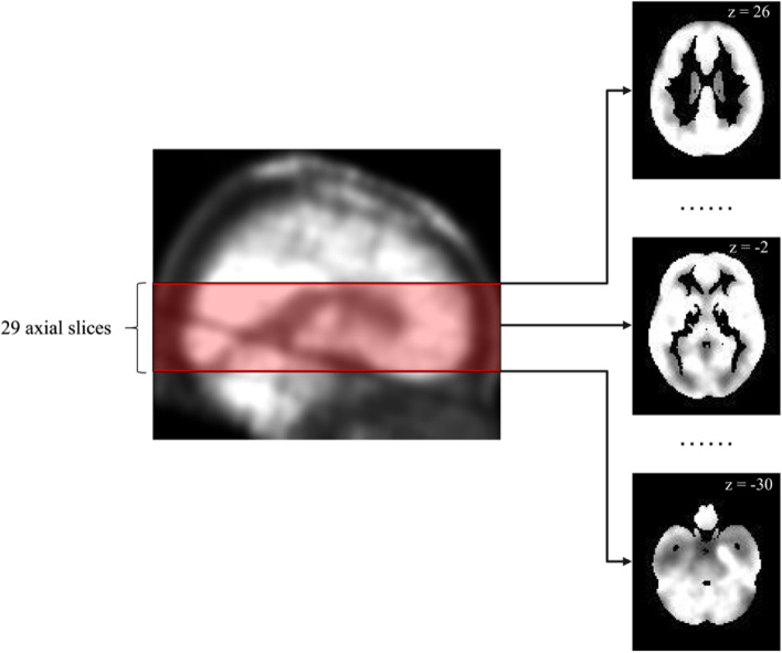
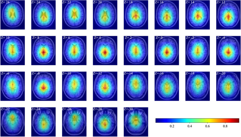
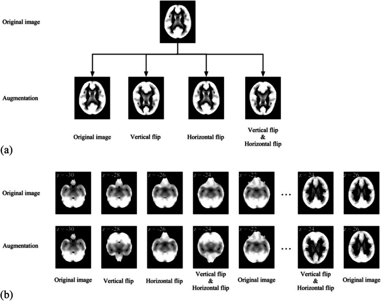
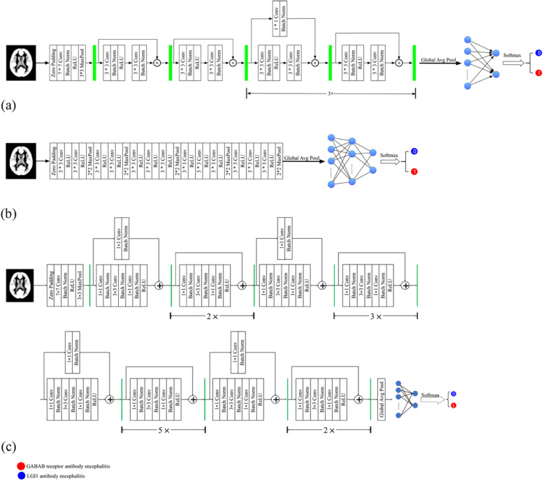
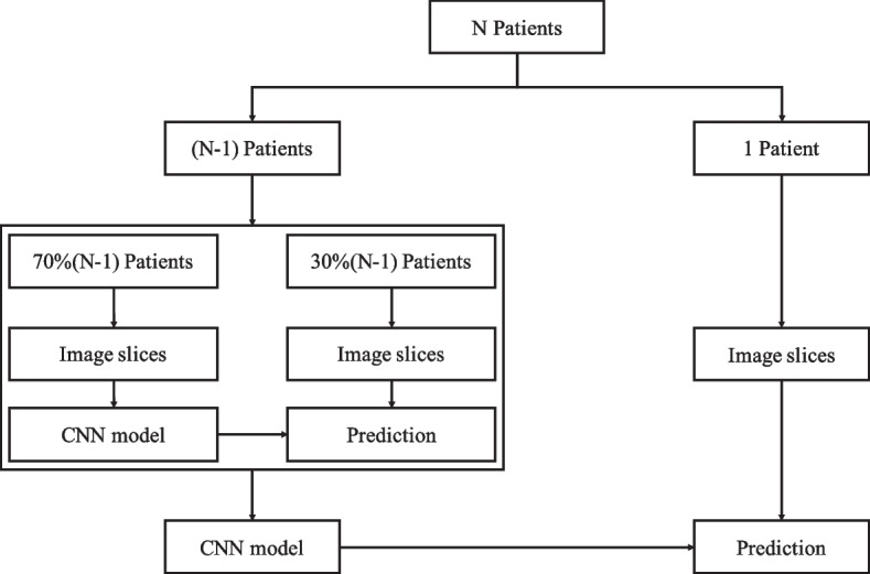
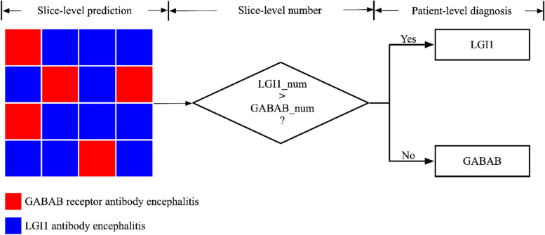
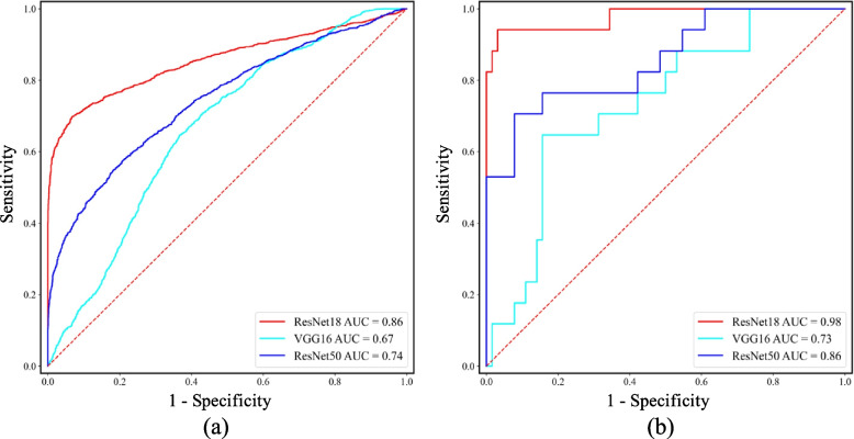
This study aims to discriminate between leucine-rich glioma-inactivated 1 (LGI1) antibody encephalitis and gamma-aminobutyric acid B (GABAB) receptor antibody encephalitis using a convolutional neural network (CNN) model. A total of 81 patients were recruited for this study. ResNet18, VGG16, and ResNet50 were trained and tested separately using 3828 positron emission tomography image slices that contained the medial temporal lobe (MTL) or basal ganglia (BG). Leave-one-out cross-validation at the patient level was used to evaluate the CNN models. The receiver operating characteristic (ROC) curve and the area under the ROC curve (AUC) were generated to evaluate the CNN models. Based on the prediction results at slice level, a decision strategy was employed to evaluate the CNN models' performance at patient level. The ResNet18 model achieved the best performance at the slice (AUC = 0.86, accuracy = 80.28%) and patient levels (AUC = 0.98, accuracy = 96.30%). Specifically, at the slice level, 73.28% (1445/1972) of image slices with GABAB receptor antibody encephalitis and 87.72% (1628/1856) of image slices with LGI1 antibody encephalitis were accurately detected. At the patient level, 94.12% (16/17) of patients with GABAB receptor antibody encephalitis and 96.88% (62/64) of patients with LGI1 antibody encephalitis were accurately detected. Heatmaps of the image slices extracted using gradient-weighted class activation mapping indicated that the model focused on the MTL and BG for classification. In general, the ResNet18 model is a potential approach for discriminating between LGI1 and GABAB receptor antibody encephalitis. Metabolism in the MTL and BG is important for discriminating between these two encephalitis subtypes.
本研究旨在使用卷积神经网络(CNN)模型区分富含亮氨酸的胶质瘤失活1(LGI1)抗体脑炎和γ-氨基丁酸B(GABAB)受体抗体脑炎。本研究共招募了81名患者。使用包含内侧颞叶(MTL)或基底神经节(BG)的3828张正电子发射断层扫描图像切片分别对ResNet18、VGG16和ResNet50进行训练和测试。在患者层面采用留一法交叉验证来评估CNN模型。生成受试者工作特征(ROC)曲线和ROC曲线下面积(AUC)以评估CNN模型。基于切片层面的预测结果,采用一种决策策略来评估CNN模型在患者层面的性能。ResNet18模型在切片(AUC = 0.86,准确率 = 80.28%)和患者层面(AUC = 0.98,准确率 = 96.30%)均取得了最佳性能。具体而言,在切片层面,准确检测出73.28%(1445/1972)的GABAB受体抗体脑炎图像切片和87.72%(1628/1856)的LGI1抗体脑炎图像切片。在患者层面,准确检测出94.12%(16/17)的GABAB受体抗体脑炎患者和96.88%(6/64)的LGI1抗体脑炎患者。使用梯度加权类激活映射提取的图像切片热图表明,该模型在分类时聚焦于MTL和BG。总体而言,ResNet18模型是区分LGI1和GABAB受体抗体脑炎的一种潜在方法。MTL和BG中的代谢对于区分这两种脑炎亚型很重要。