Institute of Immunology and Infection Research, University of Edinburgh, Edinburgh, United Kingdom.
Insitute of Microbiology, Universidad San Francisco de Quito, Quito, Ecuador.
J Clin Invest. 2023 Oct 16;133(20):e152463. doi: 10.1172/JCI152463.
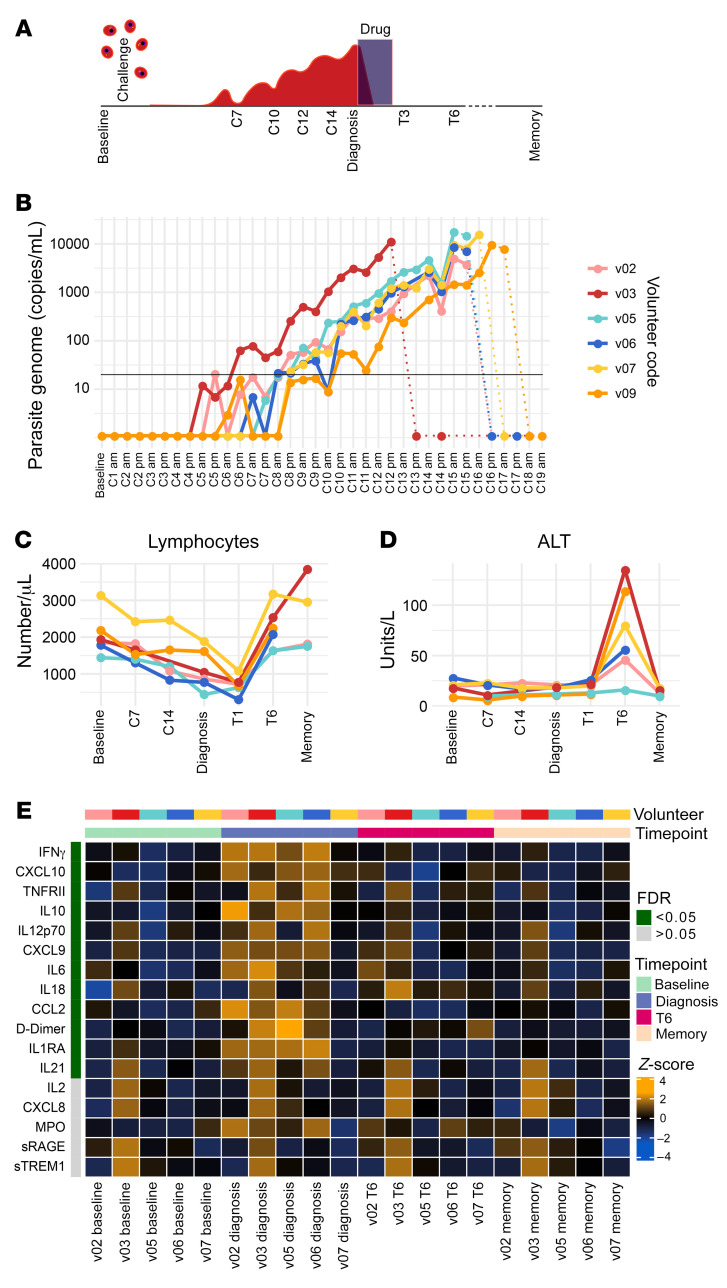
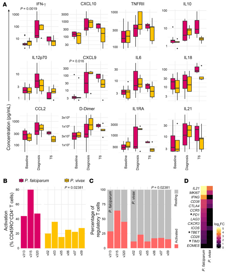
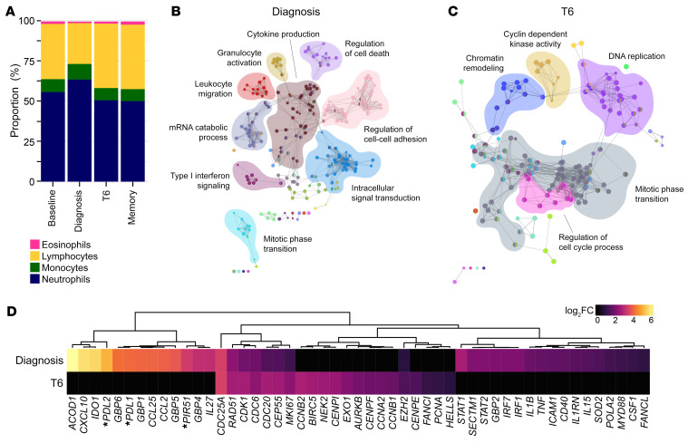
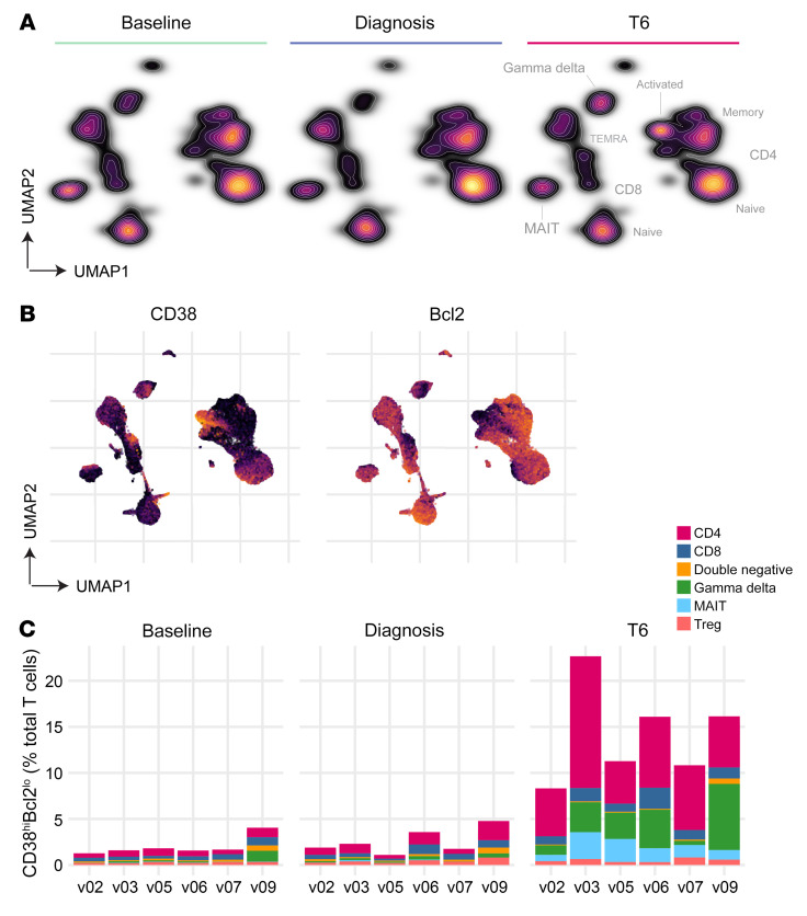
BACKGROUNDThe biology of Plasmodium vivax is markedly different from that of P. falciparum; how this shapes the immune response to infection remains unclear. To address this shortfall, we inoculated human volunteers with a clonal field isolate of P. vivax and tracked their response through infection and convalescence.METHODSParticipants were injected intravenously with blood-stage parasites and infection dynamics were tracked in real time by quantitative PCR. Whole blood samples were used for high dimensional protein analysis, RNA sequencing, and cytometry by time of flight, and temporal changes in the host response to P. vivax were quantified by linear regression. Comparative analyses with P. falciparum were then undertaken using analogous data sets derived from prior controlled human malaria infection studies.RESULTSP. vivax rapidly induced a type I inflammatory response that coincided with hallmark features of clinical malaria. This acute-phase response shared remarkable overlap with that induced by P. falciparum but was significantly elevated (at RNA and protein levels), leading to an increased incidence of pyrexia. In contrast, T cell activation and terminal differentiation were significantly increased in volunteers infected with P. falciparum. Heterogeneous CD4+ T cells were found to dominate this adaptive response and phenotypic analysis revealed unexpected features normally associated with cytotoxicity and autoinflammatory disease.CONCLUSIONP. vivax triggers increased systemic interferon signaling (cf P. falciparum), which likely explains its reduced pyrogenic threshold. In contrast, P. falciparum drives T cell activation far in excess of P. vivax, which may partially explain why falciparum malaria more frequently causes severe disease.TRIAL REGISTRATIONClinicalTrials.gov NCT03797989.FUNDINGThe European Union's Horizon 2020 Research and Innovation programme, the Wellcome Trust, and the Royal Society.
间日疟原虫的生物学特性明显不同于恶性疟原虫;这如何影响感染后的免疫反应尚不清楚。为了解决这一不足,我们用人源化的间日疟原虫野外分离株对志愿者进行了接种,并在感染和康复期间对其反应进行了跟踪。
参与者静脉内注射血期寄生虫,通过实时定量 PCR 跟踪感染动力学。全血样本用于高维蛋白分析、RNA 测序和飞行时间细胞术,通过线性回归量化宿主对间日疟原虫的反应随时间的变化。然后,使用先前控制人体疟疾感染研究中获得的类似数据集,对间日疟原虫和恶性疟原虫进行了比较分析。
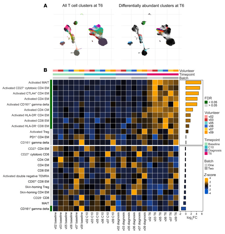
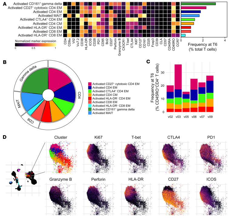
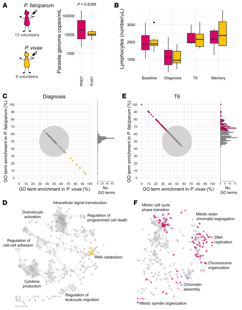
间日疟原虫迅速诱导了 I 型炎症反应,与临床疟疾的标志性特征同时发生。这种急性期反应与恶性疟原虫诱导的反应有显著重叠,但显著升高(在 RNA 和蛋白水平),导致发热的发生率增加。相比之下,感染恶性疟原虫的志愿者中 T 细胞活化和终末分化显著增加。发现异质性 CD4+T 细胞主导了这种适应性反应,表型分析揭示了通常与细胞毒性和自身炎症性疾病相关的意外特征。
间日疟原虫触发了系统性干扰素信号的增加(与恶性疟原虫相比),这可能解释了其较低的发热阈值。相比之下,恶性疟原虫诱导的 T 细胞活化远远超过间日疟原虫,这可能部分解释了为什么恶性疟原虫疟疾更常导致严重疾病。
ClinicalTrials.gov NCT03797989。
欧盟地平线 2020 研究与创新计划、惠康信托基金会和英国皇家学会。