Wylęgała Adam, Wozniak Przemysław, Sędziak-Marcinek Bogumiła, Bolek Bartłomiej, Szkodny Dominika, Wylęgała Edward
Health Promotion and Obesity Management, Pathophysiology Department, Medical University of Silesia in Katowice, 40-752 Katowice, Poland.
Chair and Clinical Department of Ophthalmology, School of Medicine in Zabrze, Medical University of Silesia in Katowice, District Railway Hospital, 40-760 Katowice, Poland.
Diagnostics (Basel). 2023 Sep 2;13(17):2846. doi: 10.3390/diagnostics13172846.
Retro-mode is a novel technique capable of creating pseudo-3D images of the retina. However, its clinical utility remains unknown. This study aimed to evaluate the Nidek Mirante multimodal imaging platform for ocular assessment in patients with various retinal conditions.
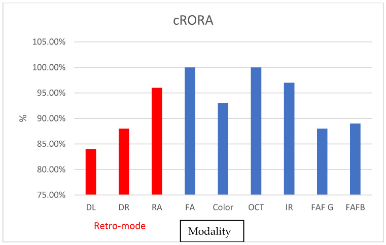
A total of 115 participants with central serous chorioretinopathy (CSR) and age-related macular degeneration (AMD) were included. Two experienced graders independently evaluated the images, and statistical analysis was performed to assess interclass correlation coefficients (ICC) between graders and modalities; Results: For CSR detection, retro-mode demonstrated exceptionally high ICC rates (ICC = 1; 100%), while color and autofluorescence (FAF) showed moderate coefficients (0.69 and 0.78, respectively). The detection of pigment epithelial detachment was high across all methods, with only retro-mode deviated right (DR) allowing detection in 69% of cases, while retro-mode DR and deviated left (DL) achieved 100% detection. FAF-green achieved a 95% detection rate. In detecting retinal atrophy, most modalities demonstrated high detection rates, with the lowest detection rates offered by retro-mode DL (ICC = 0.85) and DR (ICC = 0.89), while retro-mode ring aperture offered 0.97. Infra-red and fluorescein angiography imaging offered the highest detection rates among the tested modalities, with 97% and 100%, respectively.
Retro-mode showed promise for comprehensive ocular evaluation and diagnosis, with certain imaging modalities demonstrating higher accuracy in detecting specific retinal features.
后视模式是一种能够创建视网膜伪三维图像的新技术。然而,其临床实用性尚不清楚。本研究旨在评估尼德克Mirante多模态成像平台在各种视网膜疾病患者眼部评估中的应用。
共纳入115例中心性浆液性脉络膜视网膜病变(CSR)和年龄相关性黄斑变性(AMD)患者。两名经验丰富的分级人员独立评估图像,并进行统计分析以评估分级人员之间以及不同模式之间的组内相关系数(ICC)。
对于CSR检测,后视模式显示出极高的ICC率(ICC = 1;100%),而彩色和自发荧光(FAF)显示出中等系数(分别为0.69和0.78)。所有方法对色素上皮脱离的检测率都很高,只有后视模式右偏(DR)在69%的病例中能够检测到,而后视模式DR和左偏(DL)的检测率达到100%。FAF-绿色的检测率达到95%。在检测视网膜萎缩时,大多数模式显示出较高的检测率,后视模式DL(ICC = 0.85)和DR(ICC = 0.89)的检测率最低,而后视模式环形光圈的检测率为0.97。在测试的模式中,红外和荧光素血管造影成像的检测率最高,分别为97%和100%。
后视模式在全面眼部评估和诊断方面显示出前景某些成像模式在检测特定视网膜特征方面表现出更高的准确性。